New ads.

Showing posts with label protein. Show all posts

Friday, January 24, 2020

Researchers regrow damaged nerves with polymer and protein

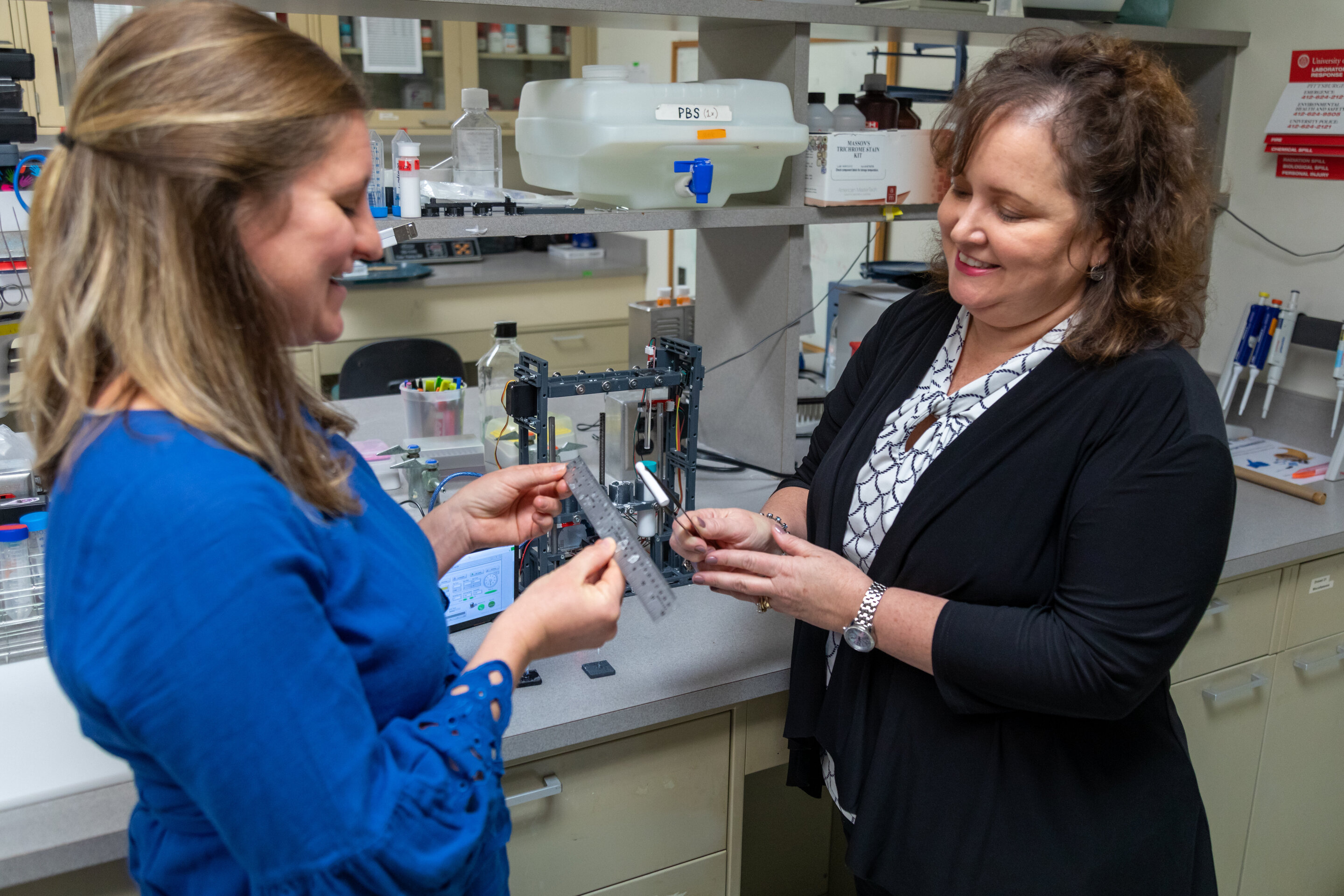
Image result for University of Pittsburgh School of Medicine researchers have created a biodegradable nerve guide -- a polymer tube -- filled with growth-promoting protein that can regenerate long sections of damaged nerves, without the need for transplanting stem cells or a donor nerve. So far, the technology has been tested in monkeys, and the results of those experiments appeared today in Science Translational Medicine. "We're the first to show a nerve guide without any cells was able to bridge a large, 2-inch gap between the nerve stump and its target muscle," said senior author Kacey Marra, Ph.D., professor of plastic surgery at Pitt and core faculty at the McGowan Institute for Regenerative Medicine. "Our guide was comparable to, and in some ways better than, a nerve graft." Half of wounded American soldiers return home with injuries to their arms and legs, which aren't well protected by body armor, often resulting in damaged nerves and disability. Among civilians, car crashes, machinery accidents, cancer treatment, diabetes and even birth trauma can cause significant nerve damage, affecting more than 20 million Americans. Peripheral nerves can regrow up to a third of an inch on their own, but if the damaged section is longer than that, the nerve can't find its target. Often, the disoriented nerve gets knotted into a painful ball called a neuroma. The most common treatment for longer segments of nerve damage is to remove a skinny sensory nerve at the back of the leg -- which causes numbness in the leg and other complications, but has the least chance of being missed -- chop it into thirds, bundle the pieces together and then sew them to the end of the damaged motor nerve, usually in the arm. But only about 40 to 60% of the motor function typically returns. "It's like you're replacing a piece of linguini with a bundle of angel hair pasta," Marra said. "It just doesn't work as well." Marra's nerve guide returned about 80% of fine motor control in the thumbs of four monkeys, each with a 2-inch nerve gap in the forearm. The guide is made of the same material as dissolvable sutures and peppered with a growth-promoting protein -- the same one delivered to the brain in a recent Parkinson's trial -- which releases slowly over the course of months. The experiment had two controls: an empty polymer tube and a nerve graft. Since monkeys' legs are relatively short, the usual clinical procedure of removing and dicing a leg nerve wouldn't work. So, the scientists removed a 2-inch segment of nerve from the forearm, flipped it around and sewed it into place, replacing linguini with linguini, and setting a high bar for the nerve guide to match. Functional recovery was just as good with Marra's guide as it was with this best-case-scenario graft, and the guide outperformed the graft when it came to restoring nerve conduction and replenishing Schwann cells -- the insulating layer around nerves that boosts electrical signals and supports regeneration. In both scenarios, it took a year for the nerve to regrow. The empty guide performed significantly worse all around. With these promising results in monkeys, Marra wants to bring her nerve guide to human patients. She's working with the Food and Drug Administration (FDA) on a first-in-human clinical trial and spinning out a startup company, AxoMax Technologies Inc. "There are no hollow tubes on the market that are approved by the FDA for nerve gaps greater than an inch. Once you get past that, no off-the-shelf tube has been shown to work," Marra said. "That's what's amazing here." Story Source: Materials provided by University of Pittsburgh. Note: Content may be edited for style and length.
Researchers regrow damaged nerves with polymer and protein
University of Pittsburgh School of Medicine researchers have created a biodegradable nerve guide -- a polymer tube -- filled with growth-promoting protein that can regenerate long sections of damaged nerves, without the need for transplanting stem cells or a donor nerve.
So far, the technology has been tested in monkeys, and the results of those experiments appeared today in Science Translational Medicine.
"We're the first to show a nerve guide without any cells was able to bridge a large, 2-inch gap between the nerve stump and its target muscle," said senior author Kacey Marra, Ph.D., professor of plastic surgery at Pitt and core faculty at the McGowan Institute for Regenerative Medicine. "Our guide was comparable to, and in some ways better than, a nerve graft."
Half of wounded American soldiers return home with injuries to their arms and legs, which aren't well protected by body armor, often resulting in damaged nerves and disability. Among civilians, car crashes, machinery accidents, cancer treatment, diabetes and even birth trauma can cause significant nerve damage, affecting more than 20 million Americans.
Peripheral nerves can regrow up to a third of an inch on their own, but if the damaged section is longer than that, the nerve can't find its target. Often, the disoriented nerve gets knotted into a painful ball called a neuroma.
The most common treatment for longer segments of nerve damage is to remove a skinny sensory nerve at the back of the leg -- which causes numbness in the leg and other complications, but has the least chance of being missed -- chop it into thirds, bundle the pieces together and then sew them to the end of the damaged motor nerve, usually in the arm. But only about 40 to 60% of the motor function typically returns.
"It's like you're replacing a piece of linguini with a bundle of angel hair pasta," Marra said. "It just doesn't work as well."
Marra's nerve guide returned about 80% of fine motor control in the thumbs of four monkeys, each with a 2-inch nerve gap in the forearm.
The guide is made of the same material as dissolvable sutures and peppered with a growth-promoting protein -- the same one delivered to the brain in a recent Parkinson's trial -- which releases slowly over the course of months.
The experiment had two controls: an empty polymer tube and a nerve graft. Since monkeys' legs are relatively short, the usual clinical procedure of removing and dicing a leg nerve wouldn't work. So, the scientists removed a 2-inch segment of nerve from the forearm, flipped it around and sewed it into place, replacing linguini with linguini, and setting a high bar for the nerve guide to match.
Functional recovery was just as good with Marra's guide as it was with this best-case-scenario graft, and the guide outperformed the graft when it came to restoring nerve conduction and replenishing Schwann cells -- the insulating layer around nerves that boosts electrical signals and supports regeneration. In both scenarios, it took a year for the nerve to regrow. The empty guide performed significantly worse all around.
With these promising results in monkeys, Marra wants to bring her nerve guide to human patients. She's working with the Food and Drug Administration (FDA) on a first-in-human clinical trial and spinning out a startup company, AxoMax Technologies Inc.
"There are no hollow tubes on the market that are approved by the FDA for nerve gaps greater than an inch. Once you get past that, no off-the-shelf tube has been shown to work," Marra said. "That's what's amazing here."

Story Source:
Materials provided by University of PittsburghNote: Content may be edited for style and length.

Saturday, December 7, 2019

By targeting flu-enabling protein, antibody may protect against wide-ranging strains

Influenza virus illustration

A nationwide team of researchers has found an antibody that protects mice against a wide range of potentially lethal influenza viruses, advancing efforts to design of a universal vaccine that could either treat or protect people against all strains of the virus.
The study, which Scripps Research conducted jointly with Washington University School of Medicine in St. Louis and Icahn School of Medicine at Mount Sinai in New York, points to a new approach to tackle severe cases of the flu, including pandemics. The research is published in the Oct. 25 issue of Science.
Scripps Research's Ian Wilson, DPhil, one of three senior co-authors, says the antibody at the center of the study binds to a protein called neuraminidase, which is essential for the flu virus to replicate in the body.
The protein, located on the surface of the virus, enables infected host cells to release the virus so it can spread to other cells. Tamiflu, the most widely used drug for severe flu infection, works by inactivating neuraminidase. However, many forms of neuraminidase exist, depending on the flu strain, and such drugs aren't always effective -- particularly as resistance to the drugs is developing.
"There are many strains of influenza virus that circulate so every year we have to design and produce a new vaccine to match the most common strains of that year," says co-senior author Ali Ellebedy, PhD, an assistant professor of pathology and immunology at Washington University. "Now imagine if we could have one vaccine that protected against all influenza strains, including human, swine and other highly lethal avian influenza viruses. This antibody could be the key to design of a truly universal vaccine."
Ellebedy discovered the antibody -- an immune molecule that recognizes and attaches to a foreign molecule -- in blood taken from a patient hospitalized with flu at Barnes-Jewish Hospital in St. Louis in the winter of 2017.
Ellebedy was working on a study analyzing the immune response to flu infection in humans in collaboration with the Washington University Emergency Care and Research Core, which was sending him blood samples from consenting flu patients. He quickly noticed that a particular blood sample was unusual: In addition to containing antibodies against hemagglutinin, the major protein on the surface of the virus, it contained other antibodies that were clearly targeting something else.
"At the time we were just starting, and I was setting up my lab so we didn't have the tools to look at what else the antibodies could be targeting," says Ellebedy, an assistant professor of medicine and of molecular microbiology.
He sent three of the antibodies to co-senior author Florian Krammer, PhD, a microbiology professor at the Icahn School of Medicine at Mount Sinai. An expert on neuraminidase, Krammer tested the antibodies against his extensive library of neuraminidase proteins. At least one of the three antibodies blocked neuraminidase activity in all known types of neuraminidase in flu viruses, representing a variety of human and nonhuman strains.
"The breadth of the antibodies really came as a surprise to us," says Krammer. "Typically, anti-neuraminidase antibodies can be broad within a subtype, like H1N1, but an antibody with potent activity across subtypes was unheard of. At first, we did not believe our results. Especially the ability of the antibodies to cross between influenza A and influenza B viruses is just mind-boggling. It is amazing what the human immune system is capable of if presented with the right antigens."
To find out whether the antibodies could be used to treat severe cases of flu, Krammer and colleagues tested them in mice that were given a lethal dose of influenza virus. All three antibodies were effective against many strains, and one antibody, called "1G01," protected against all 12 strains tested, which included all three groups of human flu virus as well as avian and other nonhuman strains.
"All the mice survived, even if they were given the antibody 72 hours after infection," Ellebedy says. "They definitely got sick and lost weight, but we still saved them. It was remarkable. It made us think that you might be able to use this antibody in an intensive care scenario when you have someone sick with flu and it's too late to use Tamiflu."
Tamiflu must be administered within 24 hours of symptoms. A drug that could be used later would help many people diagnosed after the Tamiflu window has closed. But before the researchers could even think of designing such a drug based on the antibody, they needed to understand how it was interfering with neuraminidase.
They turned to Scripps Research's Wilson, known globally for his work as a structural biologist. Wilson is Chair of the Institute's Department of Integrative Structural and Computational Biology, and has made numerous seminal findings that have shaped efforts to develop universal vaccines for flu and other complex viruses such as HIV.
Wilson and Xueyong Zhu, PhD, a staff scientist in Wilson's lab, mapped the structures of the antibodies while they were bound to neuraminidase. They found that the antibodies each had a loop that slid inside the active site of neuraminidase like a stick between gears. The loops prevented neuraminidase from releasing new virus particles from the surface of cells, thereby breaking the cycle of viral production in host cells.
"We were surprised at how these antibodies managed to insert a single loop into the conserved active site without contacting the surrounding hypervariable regions, thereby achieving much greater breadth against the neuraminidase of different influenza viruses than we have seen before," Wilson says.
The structures showed that the antibodies provide such broad protection because they target the conserved residues in the active site of the neuraminidase protein. That site stays much the same across distantly related flu strains because even minor changes could abolish the protein's ability to do its job, thereby preventing the virus from replicating.
The researchers are working on developing new and improved treatments and vaccines for influenza based on antibody 1G01.
"Neuraminidase has been ignored as a vaccine candidate for a long time," Ellebedy says. "These antibodies tell us that it should not have been overlooked. Now that we know what a broadly protective antibody to the neuraminidase looks like, we have an alternative approach to start designing novel vaccines that induce antibodies like this. And that could be really important if we are going to figure out how to design a truly universal vaccine."
The study was supported by the grants from the National Institute of Allergy and Infectious Diseases (R01 AI117287, R21 AI139813, U01 AI141990) and the National Institutes of Health (R56 AI117675).

Story Source:
Materials provided by Scripps Research InstituteNote: Content may be edited for style and length.

Saturday, November 23, 2019

Little-known protein appears to play important role in obesity and metabolic disease

Obese related protein structure

With unexpected findings about a protein that's highly expressed in fat tissue, scientists at Scripps Research have opened the door to critical new understandings about obesity and metabolism. Their discovery, which appears Nov. 20 in the journal Nature, could lead to new approaches for addressing obesity and potentially many other diseases.
The signaling protein, known as PGRMC2, had not been extensively studied in the past. Short for "progesterone receptor membrane component 2," it had been detected in the uterus, liver and several areas of the body. But the lab of Enrique Saez, PhD, saw that it was most abundant in fat tissue -- particularly in brown fat, which turns food into heat to maintain body temperature -- and became interested in its function there.
An important role: heme's travel guide
The team built on their recent discovery that PGRMC2 binds to and releases an essential molecule called heme. Recently in the spotlight for its role in providing flavor to the plant-based Impossible Burger, heme holds a much more significant role in the body. The iron-containing molecule travels within cells to enable crucial life processes such as cellular respiration, cell proliferation, cell death and circadian rhythms.
Using biochemical techniques and advanced assays in cells, Saez and his team found that PGRMC2 is a "chaperone" of heme, encapsulating the molecule and transporting it from the cell's mitochondria, where heme is created, to the nucleus, where it helps carry out important functions. Without a protective chaperone, heme would react with -- and destroy -- everything in its path.
"Heme's significance to many cellular processes has been known for a long time," says Saez, associate professor in the Department of Molecular Medicine. "But we also knew that heme is toxic to the cellular materials around it and would need some sort of shuttling pathway. Until now, there were many hypotheses, but the proteins that traffic heme had not been identified."
An innovative approach for obesity?
Through studies involving mice, the scientists established PGRMC2 as the first intracellular heme chaperone to be described in mammals. However, they didn't stop there; they sought to find out what happens in the body if this protein doesn't exist to transport heme.
And that's how they made their next big discovery: Without PGRMC2 present in their fat tissues, mice that were fed a high-fat diet became intolerant to glucose and insensitive to insulin -- hallmark symptoms of diabetes and other metabolic diseases. By contrast, obese-diabetic mice that were treated with a drug to activate PGRMC2 function showed a substantial improvement of symptoms associated with diabetes.
"We saw the mice get better, becoming more glucose tolerant and less resistant to insulin," Saez says. "Our findings suggest that modulating PGRMC2 activity in fat tissue may be a useful pharmacological approach for reverting some of the serious health effects of obesity."
The team also evaluated how the protein changes other functions of brown and white fat, says the study's lead author, Andrea Galmozzi, PhD. "The first surprise finding was that the brown fat looked white," he says.
Brown fat, which is normally the highest in heme content, is often considered the "good fat." One of its key roles is to generate heat to maintain body temperature. Among mice that were unable to produce PGRMC2 in their fat tissues, temperatures dropped quickly when placed in a cold environment.
"Even though their brain was sending the right signals to turn on the heat, the mice were unable to defend their body temperature," Galmozzi says. "Without heme, you get mitochondrial dysfunction and the cell has no means to burn energy to generate heat."
Saez believes it's possible that activating the heme chaperone in other organs -- including the liver, where a large amount of heme is made -- could help mitigate the effects of other metabolic disorders such as non-alcoholic steatohepatitis (NASH), which is a major cause of liver transplantation today.
"We're curious to know whether this protein performs the same role in other tissues where we see defects in heme that result in disease" Saez says.

Story Source:
Materials provided by Scripps Research InstituteNote: Content may be edited for style and length.

Friday, November 22, 2019

Little-known protein appears to play important role in obesity and metabolic disease

With unexpected findings about a protein that's highly expressed in fat tissue, scientists at Scripps Research have opened the door to critical new understandings about obesity and metabolism. Their discovery, which appears Nov. 20 in the journal Nature, could lead to new approaches for addressing obesity and potentially many other diseases.
The signaling protein, known as PGRMC2, had not been extensively studied in the past. Short for "progesterone receptor membrane component 2," it had been detected in the uterus, liver and several areas of the body. But the lab of Enrique Saez, PhD, saw that it was most abundant in fat tissue -- particularly in brown fat, which turns food into heat to maintain body temperature -- and became interested in its function there.
An important role: heme's travel guide
The team built on their recent discovery that PGRMC2 binds to and releases an essential molecule called heme. Recently in the spotlight for its role in providing flavor to the plant-based Impossible Burger, heme holds a much more significant role in the body. The iron-containing molecule travels within cells to enable crucial life processes such as cellular respiration, cell proliferation, cell death and circadian rhythms.
Using biochemical techniques and advanced assays in cells, Saez and his team found that PGRMC2 is a "chaperone" of heme, encapsulating the molecule and transporting it from the cell's mitochondria, where heme is created, to the nucleus, where it helps carry out important functions. Without a protective chaperone, heme would react with -- and destroy -- everything in its path.
"Heme's significance to many cellular processes has been known for a long time," says Saez, associate professor in the Department of Molecular Medicine. "But we also knew that heme is toxic to the cellular materials around it and would need some sort of shuttling pathway. Until now, there were many hypotheses, but the proteins that traffic heme had not been identified."
An innovative approach for obesity?
Through studies involving mice, the scientists established PGRMC2 as the first intracellular heme chaperone to be described in mammals. However, they didn't stop there; they sought to find out what happens in the body if this protein doesn't exist to transport heme.
And that's how they made their next big discovery: Without PGRMC2 present in their fat tissues, mice that were fed a high-fat diet became intolerant to glucose and insensitive to insulin -- hallmark symptoms of diabetes and other metabolic diseases. By contrast, obese-diabetic mice that were treated with a drug to activate PGRMC2 function showed a substantial improvement of symptoms associated with diabetes.
"We saw the mice get better, becoming more glucose tolerant and less resistant to insulin," Saez says. "Our findings suggest that modulating PGRMC2 activity in fat tissue may be a useful pharmacological approach for reverting some of the serious health effects of obesity."
The team also evaluated how the protein changes other functions of brown and white fat, says the study's lead author, Andrea Galmozzi, PhD. "The first surprise finding was that the brown fat looked white," he says.
Brown fat, which is normally the highest in heme content, is often considered the "good fat." One of its key roles is to generate heat to maintain body temperature. Among mice that were unable to produce PGRMC2 in their fat tissues, temperatures dropped quickly when placed in a cold environment.
"Even though their brain was sending the right signals to turn on the heat, the mice were unable to defend their body temperature," Galmozzi says. "Without heme, you get mitochondrial dysfunction and the cell has no means to burn energy to generate heat."
Saez believes it's possible that activating the heme chaperone in other organs -- including the liver, where a large amount of heme is made -- could help mitigate the effects of other metabolic disorders such as non-alcoholic steatohepatitis (NASH), which is a major cause of liver transplantation today.
"We're curious to know whether this protein performs the same role in other tissues where we see defects in heme that result in disease" Saez says.

Story Source:
Materials provided by Scripps Research InstituteNote: Content may be edited for style and length.