New ads.

Showing posts with label may. Show all posts

Saturday, July 11, 2020

The best material for homemade face masks may be a combination of two fabrics

The best material for homemade face masks

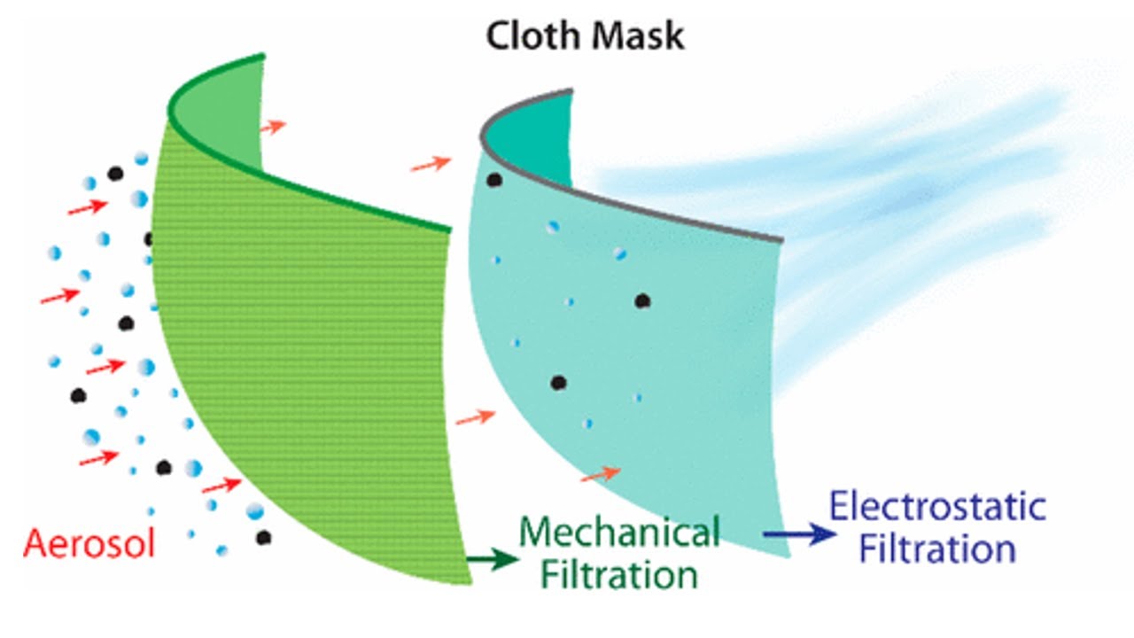
In the wake of the COVID-19 pandemic, the U.S. Centers for Disease Control and Prevention recommends that people wear masks in public. Because N95 and surgical masks are scarce and should be reserved for health care workers, many people are making their own coverings. Now, researchers report in ACS Nano that a combination of cotton with natural silk or chiffon can effectively filter out aerosol particles -- if the fit is good.
SARS-CoV-2, the new coronavirus that causes COVID-19, is thought to spread mainly through respiratory droplets when an infected person coughs, sneezes, speaks or breathes. These droplets form in a wide range of sizes, but the tiniest ones, called aerosols, can easily slip through the openings between certain cloth fibers, leading some people to question whether cloth masks can actually help prevent disease. Therefore, Supratik Guha at the University of Chicago and colleagues wanted to study the ability of common fabrics, alone or in combination, to filter out aerosols similar in size to respiratory droplets.
The researchers used an aerosol mixing chamber to produce particles ranging from 10 nm to 6 μm in diameter. A fan blew the aerosol across various cloth samples at an airflow rate corresponding to a person's respiration at rest, and the team measured the number and size of particles in air before and after passing through the fabric. One layer of a tightly woven cotton sheet combined with two layers of polyester-spandex chiffon -- a sheer fabric often used in evening gowns -- filtered out the most aerosol particles (80-99%, depending on particle size), with performance close to that of an N95 mask material. Substituting the chiffon with natural silk or flannel, or simply using a cotton quilt with cotton-polyester batting, produced similar results. The researchers point out that tightly woven fabrics, such as cotton, can act as a mechanical barrier to particles, whereas fabrics that hold a static charge, like certain types of chiffon and natural silk, serve as an electrostatic barrier. However, a 1% gap reduced the filtering efficiency of all masks by half or more, emphasizing the importance of a properly fitted mask.
The authors acknowledge use of the U.S. Department of Energy's Center for Nanoscale Materials user facility at Argonne National Laboratory and funding from the U.S. Department of Defense's Vannevar Bush Fellowship.

Story Source:
Materials provided by American Chemical SocietyNote: Content may be edited for style and length.

Journal Reference:
  1. Abhiteja Konda, Abhinav Prakash, Gregory A. Moss, Michael Schmoldt, Gregory D. Grant, Supratik Guha. Aerosol Filtration Efficiency of Common Fabrics Used in Respiratory Cloth MasksACS Nano, 2020; DOI: 10.1021/acsnano.0c03252

Wednesday, May 20, 2020

Our ability to focus may falter after eating one meal high in saturated fat

Our ability to focus may falter after eating one meal high in saturated fat

Fatty food may feel like a friend during these troubled times, but new research suggests that eating just one meal high in saturated fat can hinder our ability to concentrate -- not great news for people whose diets have gone south while they're working at home during the COVID-19 pandemic.
The study compared how 51 women performed on a test of their attention after they ate either a meal high in saturated fat or the same meal made with sunflower oil, which is high in unsaturated fat.
Their performance on the test was worse after eating the high-saturated-fat meal than after they ate the meal containing a healthier fat, signaling a link between that fatty food and the brain.
Researchers were also looking at whether a condition called leaky gut, which allows intestinal bacteria to enter the bloodstream, had any effect on concentration. Participants with leakier guts performed worse on the attention assessment no matter which meal they had eaten.
The loss of focus after a single meal was eye-opening for the researchers.
"Most prior work looking at the causative effect of the diet has looked over a period of time. And this was just one meal -- it's pretty remarkable that we saw a difference," said Annelise Madison, lead author of the study and a graduate student in clinical psychology at The Ohio State University.
Madison also noted that the meal made with sunflower oil, while low in saturated fat, still contained a lot of dietary fat.
"Because both meals were high-fat and potentially problematic, the high-saturated-fat meal's cognitive effect could be even greater if it were compared to a lower-fat meal," she said.
The study is published in the American Journal of Clinical Nutrition.
Madison works in the lab of Janice Kiecolt-Glaser, professor of psychiatry and psychology and director of the Institute for Behavioral Medicine Research at Ohio State. For this work, Madison conducted a secondary analysis of data from Kiecolt-Glaser's study assessing whether high-fat meals increased fatigue and inflammation among cancer survivors.
Women in the study completed a baseline assessment of their attention during a morning visit to the lab. The tool, called a continuous performance test, is a measure of sustained attention, concentration and reaction time based on 10 minutes of computer-based activities.
The high-fat meal followed: eggs, biscuits, turkey sausage and gravy containing 60 grams of fat, either a palmitic acid-based oil high in saturated fat or the lower-saturated-fat sunflower oil. Both meals totaled 930 calories and were designed to mimic the contents of various fast-food meals such as a Burger King double whopper with cheese or a McDonald's Big Mac and medium fries.
Five hours later, the women took the continuous performance test again. Between one and four weeks later, they repeated these steps, eating the opposite meal of what they had eaten on the first visit.
Researchers also analyzed participants' fasting baseline blood samples to determine whether they contained an inflammatory molecule that signals the presence of endotoxemia -- the toxin that escapes from the intestines and enters the bloodstream when the gut barrier is compromised.
After eating the meal high in saturated fat, all of the participating women were, on average, 11 percent less able to detect target stimuli in the attention assessment. Concentration lapses were also apparent in the women with signs of leaky gut: Their response times were more erratic and they were less able to sustain their attention during the 10-minute test.
"If the women had high levels of endotoxemia, it also wiped out the between-meal differences. They were performing poorly no matter what type of fat they ate," Madison said.
Though the study didn't determine what was going on in the brain, Madison said previous research has suggested that food high in saturated fat can drive up inflammation throughout the body, and possibly the brain. Fatty acids also can cross the blood-brain barrier.
"It could be that fatty acids are interacting with the brain directly. What it does show is the power of gut-related dysregulation," she said.
The statistical analysis accounted for other potential influences on cognition, including depressive symptoms and the participants' average dietary saturated fat consumption. The women in the study ate three standardized meals and fasted for 12 hours before each lab visit to reduce diet variations that could affect their physiological response to the high-fat meals.
The findings suggest concentration could be even more impaired in people stressed by the pandemic who are turning to fatty foods for comfort, Kiecolt-Glaser said.
"What we know is that when people are more anxious, a good subset of us will find high-saturated-fat food more enticing than broccoli," she said. "We know from other research that depression and anxiety can interfere with concentration and attention as well. When we add that on top of the high-fat meal, we could expect the real-world effects to be even larger."
This work was supported in part by the National Institutes of Health.
Additional co-authors, all from Ohio State, included Martha Belury, Rebecca Andridge, M. Rosie Shrout, Megan Renna, William Malarkey and Michael Bailey.

Story Source:
Materials provided by Ohio State University. Original written by Emily Caldwell. Note: Content may be edited for style and length.

Journal Reference:
  1. Janice K Kiecolt-Glaser, Michael T Bailey, William B Malarkey, Megan E Renna, M Rosie Shrout, Rebecca Andridge, Martha A Belury, Annelise A Madison. Afternoon distraction: a high-saturated-fat meal and endotoxemia impact postmeal attention in a randomized crossover trialThe American Journal of Clinical Nutrition, 2020; DOI: 10.1093/ajcn/nqaa085

Tuesday, December 24, 2019

Early-life exposure to dogs may lessen risk of developing schizophrenia

Dog and chalkboard addition (stock image).

Dogs spontaneously process basic numerical quantities, using a distinct part of their brains that corresponds closely to number-responsive neural regions in humans, finds a study at Emory University.
Biology Letters published the results, which suggest that a common neural mechanism has been deeply conserved across mammalian evolution.
"Our work not only shows that dogs use a similar part of their brain to process numbers of objects as humans do -- it shows that they don't need to be trained to do it," says Gregory Berns, Emory professor of psychology and senior author of the study.
"Understanding neural mechanisms -- both in humans and across species -- gives us insights into both how our brains evolved over time and how they function now," says co-author Stella Lourenco, an associate professor of psychology at Emory.
Such insights, Lourenco adds, may one day lead to practical applications such as treating brain abnormalities and improving artificial intelligence systems.
Lauren Aulet, a PhD candidate in Lourenco's lab, is first author of the study.
The study used functional magnetic resonance imaging (fMRI) to scan dogs' brains as they viewed varying numbers of dots flashed on a screen. The results showed that the dogs' parietotemporal cortex responded to differences in the number of the dots. The researchers held the total area of the dots constant, demonstrating that it was the number of the dots, not the size, that generated the response.
The approximate number system supports the ability to rapidly estimate a quantity of objects in a scene, such as the number of predators approaching or the amount of food available for foraging. Evidence suggests that humans primarily draw on their parietal cortex for this ability, which is present even in infancy.
This basic sensitivity to numerical information, known as numerosity, does not rely on symbolic thought or training and appears to be widespread throughout the animal kingdom. Much of the research in non-humans, however, has involved intensive training of the subjects.
Previous research, for example, has found that particular neurons in the parietal cortex of monkeys are attuned to numerical values. Such studies had not clarified whether numerosity is a spontaneous system in non-human primates, because the subjects underwent many trials and received rewards for selecting scenes with greater numbers of dots in preparation for the experiments.
Behavioral studies in dogs that were trained in the task of discriminating between different quantities of objects have also indicated that dogs are sensitive to numerosity.
The Emory researchers wanted to delve further into the neural underpinnings of canine number perception using fMRI.
Berns is founder of the Dog Project, which is researching evolutionary questions surrounding man's best, and oldest friend. The project was the first to train dogs to voluntarily enter an fMRI scanner and remain motionless during scanning, without restraint or sedation.
Lourenco primarily researches human visual perception, cognition and development.
Eleven dogs of varying breeds were involved in the current fMRI experiments. The dogs did not receive advance training in numerosity. After entering the fMRI, they passively viewed dot arrays that varied in numerical value. Eight of the 11 dogs showed greater activation in the parietotemporal cortex when the ratio between alternating dot arrays was more dissimilar than when the numerical values were constant.
"We went right to the source, observing the dogs' brains, to get a direct understanding of what their neurons were doing when the dogs viewed varying quantities of dots," Aulet says. "That allowed us to bypass the weaknesses of previous behavioral studies of dogs and some other species."
Humans and dogs are separated by 80 million years of evolution, Berns notes. "Our results provide some of the strongest evidence yet that numerosity is a shared neural mechanism that goes back at least that far," he says.
Unlike dogs and other animals, humans are able to build on basic numerosity in order to do more complex math, drawing primarily on the prefrontal cortex. "Part of the reason that we are able to do calculus and algebra is because we have this fundamental ability for numerosity that we share with other animals," Aulet says. "I'm interested in learning how we evolved that higher math ability and how these skills develop over time in individuals, starting with basic numerosity in infancy."
Additional authors of the study include Veronica Chiu and Ashley Prichard, Emory graduate students in psychology, and Mark Spivak, CEO of Comprehensive Pet Therapy. Spivak and Berns co-founded Dog Star Technologies to develop techniques to study how dogs perceive the world.
The work was supported by the National Institutes of Health, the John Merck Fund and the Office of Naval Research.

Story Source:
Materials provided by Emory Health Sciences. Original written by Carol Clark. Note: Content may be edited for style and length.

NASA’s Helical Engine Design that Uses Closed-Cycle Propellant; A Proposed Stardrive that May Enable Interstellar Travel


Twentieth Century technology has relied on the use of fuels and chemical propellants to propel our ships, planes, and cars. The propulsion technology of the future will not use chemical combustion to produce thrust, and the 21st century will see the emergence of propellant-less propulsion systems. Such technologies will provide the means to travel faster than ever before at a fraction of current costs and with no pollution by-products.
This becomes absolutely crucial for interplanetary and interstellar travel, as we have stated before in RSF commentary1 reporting on Resonance-based technology may provide inertial mass reduction—the future of space travel will not be performed with chemical propellants. As an example, to date the most viable proposal for an interstellar mission with current technological capabilities is the Breakthrough Starshot project which will use a fleet of light sail probes propelled to 20% percent the speed of light via laser pulses.
Considering the significant limitations of combustion-based propulsion (as well as the harmful environmental impacts), there is a strong drive to develop the next-generation propulsion systems that will move us into the next phase of technological advancement. Torus Tech, a research and development company founded by Nassim Haramein, the founder of the Resonance Science Foundation, is researching quantum vacuum engineering technologies that will enable gravitational control and zero-point energy production.
NASA is also moving forward with developing novel propulsion systems. NASA has been working on a number of small skunkworks projects, one of which—the EmDrive—has shown promise as a viable methodology for producing thrust with microwave resonance. In a new proposal, David Burns at NASA’s Marshall Space Flight Center has described a novel propellant-less propulsion system that, much like the EmDrive, utilizes relativistic effects to generate thrust in what he calls a Relativistic Momentum Transfer Model. David Burns describes his novel design in a research paper the Helical Engine. The drive derives its appellation from the core engine which is a helical particle accelerator. The core propels ions down a helical beam guide, which imparts both a z-axis velocity component and a rotational component—it is tantamount to an ionic vortex propelled (rotating in the X and Y axes) propelled along its Z-axis. The helical beam guide is tapered, so that the base has a smaller diameter than the top, in this way ions moving through the waveguide can be accelerated at one end while the radius of rotation increases, increasing the absolute velocity; and decelerated on the return with a decreasing radius of rotation, reducing the absolute velocity.

The end result is that the ions have more mass at one end of the core where they transfer their momentum to the ship, and less mass at the other end, resulting in a net-increase of momentum in the direction of travel. The core is a closed-loop helical beam guide, so no ions are required to be added or expelled, and because of the relativistic effects the drive essentially produces thrust by expelling the ions’ mass as “propellant”. The velocity change for an ion traveling through a single cycle is small, however the beam guide contains over 3×10¹² ions that complete over a half million round trip cycles each second, in which each ion applies a force aligned with the direction of travel of the ship.
There are significant challenges before the Helical Engine can become a practical technology. It requires a lot of energy to control the electric and magnetic components of the particle accelerator and produces small amounts of thrust relative to the energy requirements. It will require solar panel power for satellite operations and nuclear power for interplanetary and interstellar travel. However, with a fission reactor the engine could produce thrust indefinitely without the need for significant refueling, enabling the craft to accelerate to extremely high speeds, and the engine will be reliable as the ions are the only moving part. Further critical thinking and design will undoubtedly bring the Helical Engine to a practical propulsion technology.
RSF In-Perspective:
1We are not going to be able to travel to the stars using chemical combustion engines. In fact, interplanetary transit within our local solar system is impractical utilizing chemical combustion. Even the height of explosion-based “blow-it up” technology, external nuclear pulse propulsion, detailed in the Orion Project, would take 44 years traveling at its top speed (0.1c) to reach the nearest stellar system to our own. The reasons for becoming an interstellar civilization are myriad and a prerequisite to the long-term survivability of humanity. In addition, the same technology that would get us to the stars would enable us to travel on the surface of earth without producing pollutants that alter the atmosphere and environment, allow for the means to feasibly capture and sequester green-house gases, and the ability to deflect potentially devastating asteroids and meteoroids. As such, a technology with even the smallest potential to offer novel means of propulsion and energy production should be explored in earnest, even if current physics theory say it is “impossible”.
Resonance-based technology may provide inertial mass reduction. By: William Brown, Biophysicist with the Resonance Science Foundation.

Monday, December 23, 2019

Early-life exposure to dogs may lessen risk of developing schizophrenia

Child with dog (stock image).

Ever since humans domesticated the dog, the faithful, obedient and protective animal has provided its owner with companionship and emotional well-being. Now, a study from Johns Hopkins Medicine suggests that being around "man's best friend" from an early age may have a health benefit as well -- lessening the chance of developing schizophrenia as an adult.
And while Fido may help prevent that condition, the jury is still out on whether or not there's any link, positive or negative, between being raised with Fluffy the cat and later developing either schizophrenia or bipolar disorder.
"Serious psychiatric disorders have been associated with alterations in the immune system linked to environmental exposures in early life, and since household pets are often among the first things with which children have close contact, it was logical for us to explore the possibilities of a connection between the two," says Robert Yolken, M.D., chair of the Stanley Division of Pediatric Neurovirology and professor of neurovirology in pediatrics at the Johns Hopkins Children's Center, and lead author of a research paper recently posted online in the journal PLOS One.
In the study, Yolken and colleagues at Sheppard Pratt Health System in Baltimore investigated the relationship between exposure to a household pet cat or dog during the first 12 years of life and a later diagnosis of schizophrenia or bipolar disorder. For schizophrenia, the researchers were surprised to see a statistically significant decrease in the risk of a person developing the disorder if exposed to a dog early in life. Across the entire age range studied, there was no significant link between dogs and bipolar disorder, or between cats and either psychiatric disorder.
The researchers caution that more studies are needed to confirm these findings, to search for the factors behind any strongly supported links, and to more precisely define the actual risks of developing psychiatric disorders from exposing infants and children under age 13 to pet cats and dogs.
According to the American Pet Products Association's most recent National Pet Owners Survey, there are 94 million pet cats and 90 million pet dogs in the United States. Previous studies have identified early life exposures to pet cats and dogs as environmental factors that may alter the immune system through various means, including allergic responses, contact with zoonotic (animal) bacteria and viruses, changes in a home's microbiome, and pet-induced stress reduction effects on human brain chemistry.
Some investigators, Yolken notes, suspect that this "immune modulation" may alter the risk of developing psychiatric disorders to which a person is genetically or otherwise predisposed.
In their current study, Yolken and colleagues looked at a population of 1,371 men and women between the ages of 18 and 65 that consisted of 396 people with schizophrenia, 381 with bipolar disorder and 594 controls. Information documented about each person included age, gender, race/ethnicity, place of birth and highest level of parental education (as a measure of socioeconomic status). Patients with schizophrenia and bipolar disorder were recruited from inpatient, day hospital and rehabilitation programs of Sheppard Pratt Health System. Control group members were recruited from the Baltimore area and were screened to rule out any current or past psychiatric disorders.
All study participants were asked if they had a household pet cat or dog or both during their first 12 years of life. Those who reported that a pet cat or dog was in their house when they were born were considered to be exposed to that animal since birth.
The relationship between the age of first household pet exposure and psychiatric diagnosis was defined using a statistical model that produces a hazard ratio -- a measure over time of how often specific events (in this case, exposure to a household pet and development of a psychiatric disorder) happen in a study group compared to their frequency in a control group. A hazard ratio of 1 suggests no difference between groups, while a ratio greater than 1 indicates an increased likelihood of developing schizophrenia or bipolar disorder. Likewise, a ratio less than 1 shows a decreased chance.
Analyses were conducted for four age ranges: birth to 3, 4 to 5, 6 to 8 and 9 to 12.
Surprisingly, Yolken says, the findings suggests that people who are exposed to a pet dog before their 13th birthday are significantly less likely -- as much as 24% -- to be diagnosed later with schizophrenia.
"The largest apparent protective effect was found for children who had a household pet dog at birth or were first exposed after birth but before age 3," he says.
Yolken adds that if it is assumed that the hazard ratio is an accurate reflection of relative risk, then some 840,000 cases of schizophrenia (24% of the 3.5 million people diagnosed with the disorder in the United States) might be prevented by pet dog exposure or other factors associated with pet dog exposure.
"There are several plausible explanations for this possible 'protective' effect from contact with dogs -- perhaps something in the canine microbiome that gets passed to humans and bolsters the immune system against or subdues a genetic predisposition to schizophrenia," Yolken says.
For bipolar disorder, the study results suggest there is no risk association, either positive or negative, with being around dogs as an infant or young child.
Overall for all ages examined, early exposure to pet cats was neutral as the study could not link felines with either an increased or decreased risk of developing schizophrenia or bipolar disorder.
"However, we did find a slightly increased risk of developing both disorders for those who were first in contact with cats between the ages of 9 and 12," Yolken says. "This indicates that the time of exposure may be critical to whether or not it alters the risk."
One example of a suspected pet-borne trigger for schizophrenia is the disease toxoplasmosis, a condition in which cats are the primary hosts of a parasite transmitted to humans via the animals' feces. Pregnant women have been advised for years not to change cat litter boxes to eliminate the risk of the illness passing through the placenta to their fetuses and causing a miscarriage, stillbirth, or potentially, psychiatric disorders in a child born with the infection.
In a 2003 review paper, Yolken and colleague E. Fuller Torrey, M.D., associate director of research at the Stanley Medical Research Institute in Bethesda, Maryland, provided evidence from multiple epidemiological studies conducted since 1953 that showed there also is a statistical connection between a person exposed to the parasite that causes toxoplasmosis and an increased risk of developing schizophrenia. The researchers found that a large number of people in those studies who were diagnosed with serious psychiatric disorders, including schizophrenia, also had high levels of antibodies to the toxoplasmosis parasite.
Because of this finding and others like it, most research has focused on investigating a potential link between early exposure to cats and psychiatric disorder development. Yolken says the most recent study is among the first to consider contact with dogs as well.
"A better understanding of the mechanisms underlying the associations between pet exposure and psychiatric disorders would allow us to develop appropriate prevention and treatment strategies," Yolken says.
Working with Yolken on the research team are the following members from Sheppard Pratt Health System: Cassie Stallings, Andrea Origoni, Emily Katsafanas, Kevin Sweeney, Amalia Squire, and Faith Dickerson, Ph.D., M.P.H.
The study was largely supported by grants from the Stanley Medical Research Institute.
Story Source:
Materials provided by Johns Hopkins MedicineNote: Content may be edited for style and length.

Saturday, December 21, 2019

Early-life exposure to dogs may lessen risk of developing schizophrenia

Child with dog (stock image).
Credit: © melounix / Adobe Stock

Ever since humans domesticated the dog, the faithful, obedient and protective animal has provided its owner with companionship and emotional well-being. Now, a study from Johns Hopkins Medicine suggests that being around "man's best friend" from an early age may have a health benefit as well -- lessening the chance of developing schizophrenia as an adult.
And while Fido may help prevent that condition, the jury is still out on whether or not there's any link, positive or negative, between being raised with Fluffy the cat and later developing either schizophrenia or bipolar disorder.
"Serious psychiatric disorders have been associated with alterations in the immune system linked to environmental exposures in early life, and since household pets are often among the first things with which children have close contact, it was logical for us to explore the possibilities of a connection between the two," says Robert Yolken, M.D., chair of the Stanley Division of Pediatric Neurovirology and professor of neurovirology in pediatrics at the Johns Hopkins Children's Center, and lead author of a research paper recently posted online in the journal PLOS One.
In the study, Yolken and colleagues at Sheppard Pratt Health System in Baltimore investigated the relationship between exposure to a household pet cat or dog during the first 12 years of life and a later diagnosis of schizophrenia or bipolar disorder. For schizophrenia, the researchers were surprised to see a statistically significant decrease in the risk of a person developing the disorder if exposed to a dog early in life. Across the entire age range studied, there was no significant link between dogs and bipolar disorder, or between cats and either psychiatric disorder.
The researchers caution that more studies are needed to confirm these findings, to search for the factors behind any strongly supported links, and to more precisely define the actual risks of developing psychiatric disorders from exposing infants and children under age 13 to pet cats and dogs.
According to the American Pet Products Association's most recent National Pet Owners Survey, there are 94 million pet cats and 90 million pet dogs in the United States. Previous studies have identified early life exposures to pet cats and dogs as environmental factors that may alter the immune system through various means, including allergic responses, contact with zoonotic (animal) bacteria and viruses, changes in a home's microbiome, and pet-induced stress reduction effects on human brain chemistry.
Some investigators, Yolken notes, suspect that this "immune modulation" may alter the risk of developing psychiatric disorders to which a person is genetically or otherwise predisposed.
In their current study, Yolken and colleagues looked at a population of 1,371 men and women between the ages of 18 and 65 that consisted of 396 people with schizophrenia, 381 with bipolar disorder and 594 controls. Information documented about each person included age, gender, race/ethnicity, place of birth and highest level of parental education (as a measure of socioeconomic status). Patients with schizophrenia and bipolar disorder were recruited from inpatient, day hospital and rehabilitation programs of Sheppard Pratt Health System. Control group members were recruited from the Baltimore area and were screened to rule out any current or past psychiatric disorders.
All study participants were asked if they had a household pet cat or dog or both during their first 12 years of life. Those who reported that a pet cat or dog was in their house when they were born were considered to be exposed to that animal since birth.
The relationship between the age of first household pet exposure and psychiatric diagnosis was defined using a statistical model that produces a hazard ratio -- a measure over time of how often specific events (in this case, exposure to a household pet and development of a psychiatric disorder) happen in a study group compared to their frequency in a control group. A hazard ratio of 1 suggests no difference between groups, while a ratio greater than 1 indicates an increased likelihood of developing schizophrenia or bipolar disorder. Likewise, a ratio less than 1 shows a decreased chance.
Analyses were conducted for four age ranges: birth to 3, 4 to 5, 6 to 8 and 9 to 12.
Surprisingly, Yolken says, the findings suggests that people who are exposed to a pet dog before their 13th birthday are significantly less likely -- as much as 24% -- to be diagnosed later with schizophrenia.
"The largest apparent protective effect was found for children who had a household pet dog at birth or were first exposed after birth but before age 3," he says.
Yolken adds that if it is assumed that the hazard ratio is an accurate reflection of relative risk, then some 840,000 cases of schizophrenia (24% of the 3.5 million people diagnosed with the disorder in the United States) might be prevented by pet dog exposure or other factors associated with pet dog exposure.
"There are several plausible explanations for this possible 'protective' effect from contact with dogs -- perhaps something in the canine microbiome that gets passed to humans and bolsters the immune system against or subdues a genetic predisposition to schizophrenia," Yolken says.
For bipolar disorder, the study results suggest there is no risk association, either positive or negative, with being around dogs as an infant or young child.
Overall for all ages examined, early exposure to pet cats was neutral as the study could not link felines with either an increased or decreased risk of developing schizophrenia or bipolar disorder.
"However, we did find a slightly increased risk of developing both disorders for those who were first in contact with cats between the ages of 9 and 12," Yolken says. "This indicates that the time of exposure may be critical to whether or not it alters the risk."
One example of a suspected pet-borne trigger for schizophrenia is the disease toxoplasmosis, a condition in which cats are the primary hosts of a parasite transmitted to humans via the animals' feces. Pregnant women have been advised for years not to change cat litter boxes to eliminate the risk of the illness passing through the placenta to their fetuses and causing a miscarriage, stillbirth, or potentially, psychiatric disorders in a child born with the infection.
In a 2003 review paper, Yolken and colleague E. Fuller Torrey, M.D., associate director of research at the Stanley Medical Research Institute in Bethesda, Maryland, provided evidence from multiple epidemiological studies conducted since 1953 that showed there also is a statistical connection between a person exposed to the parasite that causes toxoplasmosis and an increased risk of developing schizophrenia. The researchers found that a large number of people in those studies who were diagnosed with serious psychiatric disorders, including schizophrenia, also had high levels of antibodies to the toxoplasmosis parasite.
Because of this finding and others like it, most research has focused on investigating a potential link between early exposure to cats and psychiatric disorder development. Yolken says the most recent study is among the first to consider contact with dogs as well.
"A better understanding of the mechanisms underlying the associations between pet exposure and psychiatric disorders would allow us to develop appropriate prevention and treatment strategies," Yolken says.
Working with Yolken on the research team are the following members from Sheppard Pratt Health System: Cassie Stallings, Andrea Origoni, Emily Katsafanas, Kevin Sweeney, Amalia Squire, and Faith Dickerson, Ph.D., M.P.H.
The study was largely supported by grants from the Stanley Medical Research Institute.

Story Source:
Materials provided by Johns Hopkins MedicineNote: Content may be edited for style and length.

Saturday, December 7, 2019

By targeting flu-enabling protein, antibody may protect against wide-ranging strains

Influenza virus illustration

A nationwide team of researchers has found an antibody that protects mice against a wide range of potentially lethal influenza viruses, advancing efforts to design of a universal vaccine that could either treat or protect people against all strains of the virus.
The study, which Scripps Research conducted jointly with Washington University School of Medicine in St. Louis and Icahn School of Medicine at Mount Sinai in New York, points to a new approach to tackle severe cases of the flu, including pandemics. The research is published in the Oct. 25 issue of Science.
Scripps Research's Ian Wilson, DPhil, one of three senior co-authors, says the antibody at the center of the study binds to a protein called neuraminidase, which is essential for the flu virus to replicate in the body.
The protein, located on the surface of the virus, enables infected host cells to release the virus so it can spread to other cells. Tamiflu, the most widely used drug for severe flu infection, works by inactivating neuraminidase. However, many forms of neuraminidase exist, depending on the flu strain, and such drugs aren't always effective -- particularly as resistance to the drugs is developing.
"There are many strains of influenza virus that circulate so every year we have to design and produce a new vaccine to match the most common strains of that year," says co-senior author Ali Ellebedy, PhD, an assistant professor of pathology and immunology at Washington University. "Now imagine if we could have one vaccine that protected against all influenza strains, including human, swine and other highly lethal avian influenza viruses. This antibody could be the key to design of a truly universal vaccine."
Ellebedy discovered the antibody -- an immune molecule that recognizes and attaches to a foreign molecule -- in blood taken from a patient hospitalized with flu at Barnes-Jewish Hospital in St. Louis in the winter of 2017.
Ellebedy was working on a study analyzing the immune response to flu infection in humans in collaboration with the Washington University Emergency Care and Research Core, which was sending him blood samples from consenting flu patients. He quickly noticed that a particular blood sample was unusual: In addition to containing antibodies against hemagglutinin, the major protein on the surface of the virus, it contained other antibodies that were clearly targeting something else.
"At the time we were just starting, and I was setting up my lab so we didn't have the tools to look at what else the antibodies could be targeting," says Ellebedy, an assistant professor of medicine and of molecular microbiology.
He sent three of the antibodies to co-senior author Florian Krammer, PhD, a microbiology professor at the Icahn School of Medicine at Mount Sinai. An expert on neuraminidase, Krammer tested the antibodies against his extensive library of neuraminidase proteins. At least one of the three antibodies blocked neuraminidase activity in all known types of neuraminidase in flu viruses, representing a variety of human and nonhuman strains.
"The breadth of the antibodies really came as a surprise to us," says Krammer. "Typically, anti-neuraminidase antibodies can be broad within a subtype, like H1N1, but an antibody with potent activity across subtypes was unheard of. At first, we did not believe our results. Especially the ability of the antibodies to cross between influenza A and influenza B viruses is just mind-boggling. It is amazing what the human immune system is capable of if presented with the right antigens."
To find out whether the antibodies could be used to treat severe cases of flu, Krammer and colleagues tested them in mice that were given a lethal dose of influenza virus. All three antibodies were effective against many strains, and one antibody, called "1G01," protected against all 12 strains tested, which included all three groups of human flu virus as well as avian and other nonhuman strains.
"All the mice survived, even if they were given the antibody 72 hours after infection," Ellebedy says. "They definitely got sick and lost weight, but we still saved them. It was remarkable. It made us think that you might be able to use this antibody in an intensive care scenario when you have someone sick with flu and it's too late to use Tamiflu."
Tamiflu must be administered within 24 hours of symptoms. A drug that could be used later would help many people diagnosed after the Tamiflu window has closed. But before the researchers could even think of designing such a drug based on the antibody, they needed to understand how it was interfering with neuraminidase.
They turned to Scripps Research's Wilson, known globally for his work as a structural biologist. Wilson is Chair of the Institute's Department of Integrative Structural and Computational Biology, and has made numerous seminal findings that have shaped efforts to develop universal vaccines for flu and other complex viruses such as HIV.
Wilson and Xueyong Zhu, PhD, a staff scientist in Wilson's lab, mapped the structures of the antibodies while they were bound to neuraminidase. They found that the antibodies each had a loop that slid inside the active site of neuraminidase like a stick between gears. The loops prevented neuraminidase from releasing new virus particles from the surface of cells, thereby breaking the cycle of viral production in host cells.
"We were surprised at how these antibodies managed to insert a single loop into the conserved active site without contacting the surrounding hypervariable regions, thereby achieving much greater breadth against the neuraminidase of different influenza viruses than we have seen before," Wilson says.
The structures showed that the antibodies provide such broad protection because they target the conserved residues in the active site of the neuraminidase protein. That site stays much the same across distantly related flu strains because even minor changes could abolish the protein's ability to do its job, thereby preventing the virus from replicating.
The researchers are working on developing new and improved treatments and vaccines for influenza based on antibody 1G01.
"Neuraminidase has been ignored as a vaccine candidate for a long time," Ellebedy says. "These antibodies tell us that it should not have been overlooked. Now that we know what a broadly protective antibody to the neuraminidase looks like, we have an alternative approach to start designing novel vaccines that induce antibodies like this. And that could be really important if we are going to figure out how to design a truly universal vaccine."
The study was supported by the grants from the National Institute of Allergy and Infectious Diseases (R01 AI117287, R21 AI139813, U01 AI141990) and the National Institutes of Health (R56 AI117675).

Story Source:
Materials provided by Scripps Research InstituteNote: Content may be edited for style and length.

Saturday, November 30, 2019

Babies in the womb may see more than we thought

Illustration of fetus inside womb

By the second trimester, long before a baby's eyes can see images, they can detect light.
But the light-sensitive cells in the developing retina -- the thin sheet of brain-like tissue at the back of the eye -- were thought to be simple on-off switches, presumably there to set up the 24-hour, day-night rhythms parents hope their baby will follow.
University of California, Berkeley, scientists have now found evidence that these simple cells actually talk to one another as part of an interconnected network that gives the retina more light sensitivity than once thought, and that may enhance the influence of light on behavior and brain development in unsuspected ways.
In the developing eye, perhaps 3% of ganglion cells -- the cells in the retina that send messages through the optic nerve into the brain -- are sensitive to light and, to date, researchers have found about six different subtypes that communicate with various places in the brain. Some talk to the suprachiasmatic nucleus to tune our internal clock to the day-night cycle. Others send signals to the area that makes our pupils constrict in bright light.
But others connect to surprising areas: the perihabenula, which regulates mood, and the amygdala, which deals with emotions.
In mice and monkeys, recent evidence suggests that these ganglion cells also talk with one another through electrical connections called gap junctions, implying much more complexity in immature rodent and primate eyes than imagined.
"Given the variety of these ganglion cells and that they project to many different parts of the brain, it makes me wonder whether they play a role in how the retina connects up to the brain," said Marla Feller, a UC Berkeley professor of molecular and cell biology and senior author of a paper that appeared this month in the journal Current Biology. "Maybe not for visual circuits, but for non-vision behaviors. Not only the pupillary light reflex and circadian rhythms, but possibly explaining problems like light-induced migraines, or why light therapy works for depression."
Parallel systems in developing retina
The cells, called intrinsically photosensitive retinal ganglion cells (ipRGCs), were discovered only 10 years ago, surprising those like Feller who had been studying the developing retina for nearly 20 years. She played a major role, along with her mentor, Carla Shatz of Stanford University, in showing that spontaneous electrical activity in the eye during development -- so-called retinal waves -- is critical for setting up the correct brain networks to process images later on.
Hence her interest in the ipRGCs that seemed to function in parallel with spontaneous retinal waves in the developing retina.
"We thought they (mouse pups and the human fetus) were blind at this point in development," said Feller, the Paul Licht Distinguished Professor in Biological Sciences and a member of UC Berkeley's Helen Wills Neuroscience Institute. "We thought that the ganglion cells were there in the developing eye, that they are connected to the brain, but that they were not really connected to much of the rest of the retina, at that point. Now, it turns out they are connected to each other, which was a surprising thing."
UC Berkeley graduate student Franklin Caval-Holme combined two-photon calcium imaging, whole-cell electrical recording, pharmacology and anatomical techniques to show that the six types of ipRGCs in the newborn mouse retina link up electrically, via gap junctions, to form a retinal network that the researchers found not only detects light, but responds to the intensity of the light, which can vary nearly a billionfold.
Gap junction circuits were critical for light sensitivity in some ipRGC subtypes, but not others, providing a potential avenue to determine which ipRGC subtypes provide the signal for specific non-visual behaviors that light evokes.
"Aversion to light, which pups develop very early, is intensity-dependent," suggesting that these neural circuits could be involved in light-aversion behavior, Caval-Holme said. "We don't know which of these ipRGC subtypes in the neonatal retina actually contributes to the behavior, so it will be very interesting to see what role all these different subtypes have."
The researchers also found evidence that the circuit tunes itself in a way that could adapt to the intensity of light, which probably has an important role in development, Feller said.
"In the past, people demonstrated that these light-sensitive cells are important for things like the development of the blood vessels in the retina and light entrainment of circadian rhythms, but those were kind of a light on/light off response, where you need some light or no light," she said. "This seems to argue that they are actually trying to code for many different intensities of light, encoding much more information than people had previously thought."
The research was supported by the National Institutes of Health (NIH F31EY028022-03, RO1EY019498, RO1EY013528, P30EY003176).

Story Source:
Materials provided by University of California - Berkeley. Original written by Robert Sanders. Note: Content may be edited for style and length.

The world is getting wetter, yet water may become less available for North America and Eurasia

Drips from faucet in dry environment

With climate change, plants of the future will consume more water than in the present day, leading to less water available for people living in North America and Eurasia, according to a Dartmouth-led study in Nature Geoscience. The research suggests a drier future despite anticipated precipitation increases for places like the United States and Europe, populous regions already facing water stresses.
The study challenges an expectation in climate science that plants will make the world wetter in the future. Scientists have long thought that as carbon dioxide concentrations increase in the atmosphere, plants will reduce their water consumption, leaving more freshwater available in our soils and streams. This is because as more carbon dioxide accumulates in our atmosphere plants can photosynthesize the same amount while partly closing the pores (stomata) on their leaves. Closed stomata means less plant water loss to the atmosphere, increasing water in the land. The new findings reveal that this story of plants making the land wetter is limited to the tropics and the extremely high latitudes, where freshwater availability is already high and competing demands on it are low. For much of the mid-latitudes, the study finds, projected plant responses to climate change will not make the land wetter but drier, which has massive implications for millions of people.
"Approximately 60 percent of the global water flux from the land to the atmosphere goes through plants, called transpiration. Plants are like the atmosphere's straw, dominating how water flows from the land to the atmosphere. So vegetation is a massive determinant of what water is left on land for people," explained lead author Justin S. Mankin, an assistant professor of geography at Dartmouth and adjunct research scientist at Lamont-Doherty Earth Observatory at Columbia University. "The question we're asking here is, how do the combined effects of carbon dioxide and warming change the size of that straw?"
Using climate models, the study examines how freshwater availability may be affected by projected changes in the way precipitation is divided among plants, rivers and soils. For the study, the research team used a novel accounting of this precipitation partitioning, developed earlier by Mankin and colleagues to calculate the future runoff loss to future vegetation in a warmer, carbon dioxide-enriched climate.
The new study's findings revealed how the interaction of three key effects of climate change's impacts on plants will reduce regional freshwater availability. First, as carbon dioxide increases in the atmosphere, plants require less water to photosynthesize, wetting the land. Yet, second, as the planet warms, growing seasons become longer and warmer: plants have more time to grow and consume water, drying the land. Finally, as carbon dioxide concentrations increase, plants are likely to grow more, as photosynthesis becomes amplified. For some regions, these latter two impacts, extended growing seasons and amplified photosynthesis, will outpace the closing stomata, meaning more vegetation will consume more water for a longer amount of time, drying the land. As a result, for much of the mid-latitudes, plants will leave less water in soils and streams, even if there is additional rainfall and vegetation is more efficient with its water usage. The result also underscores the importance of improving how climate models represent ecosystems and their response to climate change.
The world relies on freshwater for human consumption, agriculture, hydropower, and industry. Yet, for many places, there's a fundamental disconnect between when precipitation falls and when people use this water, as is the case with California, which gets more than half of its precipitation in the winter, but peak demands are in the summer. "Throughout the world, we engineer solutions to move water from point A to point B to overcome this spatiotemporal disconnect between water supply and its demand. Allocating water is politically contentious, capital-intensive and requires really long-term planning, all of which affects some of the most vulnerable populations. Our research shows that we can't expect plants to be a universal panacea for future water availability. So, being able to assess clearly where and why we should anticipate water availability changes to occur in the future is crucial to ensuring that we can be prepared," added Mankin.
Researchers from Lamont-Doherty Earth Observatory of Columbia University, Richard Seager, Jason E. Smerdon, Benjamin I. Cook, who is also affiliated with NASA Goddard Institute for Space Studies, and A. Park Williams, contributed to this study.

Story Source:
Materials provided by Dartmouth CollegeNote: Content may be edited for style and length.