New ads.

Showing posts with label Researchers. Show all posts

Friday, January 24, 2020

Researchers regrow damaged nerves with polymer and protein

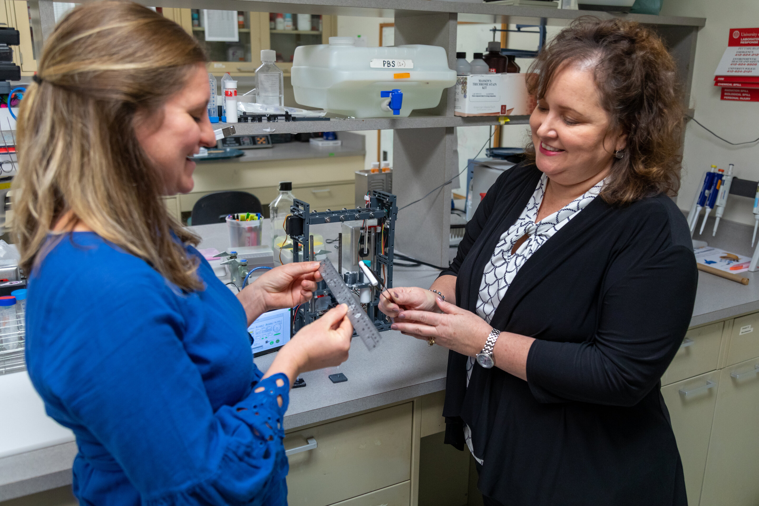
Image result for University of Pittsburgh School of Medicine researchers have created a biodegradable nerve guide -- a polymer tube -- filled with growth-promoting protein that can regenerate long sections of damaged nerves, without the need for transplanting stem cells or a donor nerve. So far, the technology has been tested in monkeys, and the results of those experiments appeared today in Science Translational Medicine. "We're the first to show a nerve guide without any cells was able to bridge a large, 2-inch gap between the nerve stump and its target muscle," said senior author Kacey Marra, Ph.D., professor of plastic surgery at Pitt and core faculty at the McGowan Institute for Regenerative Medicine. "Our guide was comparable to, and in some ways better than, a nerve graft." Half of wounded American soldiers return home with injuries to their arms and legs, which aren't well protected by body armor, often resulting in damaged nerves and disability. Among civilians, car crashes, machinery accidents, cancer treatment, diabetes and even birth trauma can cause significant nerve damage, affecting more than 20 million Americans. Peripheral nerves can regrow up to a third of an inch on their own, but if the damaged section is longer than that, the nerve can't find its target. Often, the disoriented nerve gets knotted into a painful ball called a neuroma. The most common treatment for longer segments of nerve damage is to remove a skinny sensory nerve at the back of the leg -- which causes numbness in the leg and other complications, but has the least chance of being missed -- chop it into thirds, bundle the pieces together and then sew them to the end of the damaged motor nerve, usually in the arm. But only about 40 to 60% of the motor function typically returns. "It's like you're replacing a piece of linguini with a bundle of angel hair pasta," Marra said. "It just doesn't work as well." Marra's nerve guide returned about 80% of fine motor control in the thumbs of four monkeys, each with a 2-inch nerve gap in the forearm. The guide is made of the same material as dissolvable sutures and peppered with a growth-promoting protein -- the same one delivered to the brain in a recent Parkinson's trial -- which releases slowly over the course of months. The experiment had two controls: an empty polymer tube and a nerve graft. Since monkeys' legs are relatively short, the usual clinical procedure of removing and dicing a leg nerve wouldn't work. So, the scientists removed a 2-inch segment of nerve from the forearm, flipped it around and sewed it into place, replacing linguini with linguini, and setting a high bar for the nerve guide to match. Functional recovery was just as good with Marra's guide as it was with this best-case-scenario graft, and the guide outperformed the graft when it came to restoring nerve conduction and replenishing Schwann cells -- the insulating layer around nerves that boosts electrical signals and supports regeneration. In both scenarios, it took a year for the nerve to regrow. The empty guide performed significantly worse all around. With these promising results in monkeys, Marra wants to bring her nerve guide to human patients. She's working with the Food and Drug Administration (FDA) on a first-in-human clinical trial and spinning out a startup company, AxoMax Technologies Inc. "There are no hollow tubes on the market that are approved by the FDA for nerve gaps greater than an inch. Once you get past that, no off-the-shelf tube has been shown to work," Marra said. "That's what's amazing here." Story Source: Materials provided by University of Pittsburgh. Note: Content may be edited for style and length.
Researchers regrow damaged nerves with polymer and protein
University of Pittsburgh School of Medicine researchers have created a biodegradable nerve guide -- a polymer tube -- filled with growth-promoting protein that can regenerate long sections of damaged nerves, without the need for transplanting stem cells or a donor nerve.
So far, the technology has been tested in monkeys, and the results of those experiments appeared today in Science Translational Medicine.
"We're the first to show a nerve guide without any cells was able to bridge a large, 2-inch gap between the nerve stump and its target muscle," said senior author Kacey Marra, Ph.D., professor of plastic surgery at Pitt and core faculty at the McGowan Institute for Regenerative Medicine. "Our guide was comparable to, and in some ways better than, a nerve graft."
Half of wounded American soldiers return home with injuries to their arms and legs, which aren't well protected by body armor, often resulting in damaged nerves and disability. Among civilians, car crashes, machinery accidents, cancer treatment, diabetes and even birth trauma can cause significant nerve damage, affecting more than 20 million Americans.
Peripheral nerves can regrow up to a third of an inch on their own, but if the damaged section is longer than that, the nerve can't find its target. Often, the disoriented nerve gets knotted into a painful ball called a neuroma.
The most common treatment for longer segments of nerve damage is to remove a skinny sensory nerve at the back of the leg -- which causes numbness in the leg and other complications, but has the least chance of being missed -- chop it into thirds, bundle the pieces together and then sew them to the end of the damaged motor nerve, usually in the arm. But only about 40 to 60% of the motor function typically returns.
"It's like you're replacing a piece of linguini with a bundle of angel hair pasta," Marra said. "It just doesn't work as well."
Marra's nerve guide returned about 80% of fine motor control in the thumbs of four monkeys, each with a 2-inch nerve gap in the forearm.
The guide is made of the same material as dissolvable sutures and peppered with a growth-promoting protein -- the same one delivered to the brain in a recent Parkinson's trial -- which releases slowly over the course of months.
The experiment had two controls: an empty polymer tube and a nerve graft. Since monkeys' legs are relatively short, the usual clinical procedure of removing and dicing a leg nerve wouldn't work. So, the scientists removed a 2-inch segment of nerve from the forearm, flipped it around and sewed it into place, replacing linguini with linguini, and setting a high bar for the nerve guide to match.
Functional recovery was just as good with Marra's guide as it was with this best-case-scenario graft, and the guide outperformed the graft when it came to restoring nerve conduction and replenishing Schwann cells -- the insulating layer around nerves that boosts electrical signals and supports regeneration. In both scenarios, it took a year for the nerve to regrow. The empty guide performed significantly worse all around.
With these promising results in monkeys, Marra wants to bring her nerve guide to human patients. She's working with the Food and Drug Administration (FDA) on a first-in-human clinical trial and spinning out a startup company, AxoMax Technologies Inc.
"There are no hollow tubes on the market that are approved by the FDA for nerve gaps greater than an inch. Once you get past that, no off-the-shelf tube has been shown to work," Marra said. "That's what's amazing here."

Story Source:
Materials provided by University of PittsburghNote: Content may be edited for style and length.
Wednesday, January 15, 2020

Researchers learn more about teen-age T. rex

Image result for Researchers learn more about teen-age T. rex

Researchers learn more about teen-age T. rex


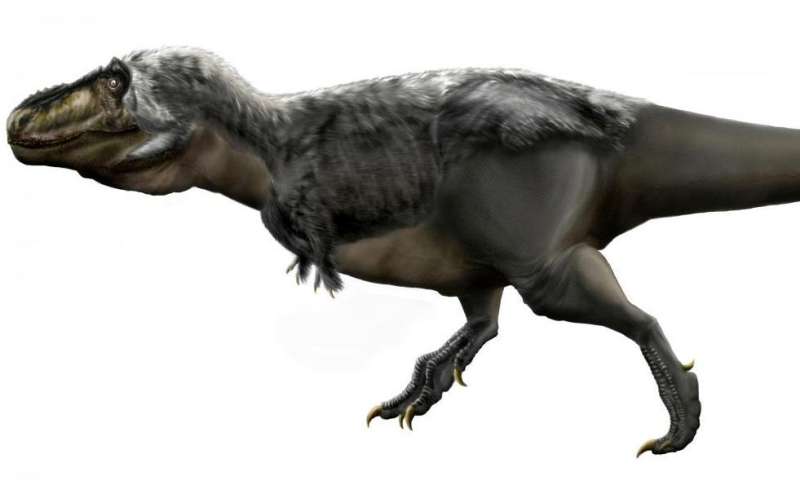
Without a doubt, Tyrannosaurus rex is the most famous dinosaur in the world. The 40-foot-long predator with bone crushing teeth inside a five-foot long head are the stuff of legend. Now, a look within the bones of two mid-sized, immature T. rex allow scientists to learn about the tyrant king's terrible teens as well.
In the early 2000s, the fossil skeletons of two comparatively small T. rex were collected from Carter County, Montana, by Burpee Museum of Natural History in Rockford, Illinois. Nicknamed "Jane" and "Petey," the tyrannosaurs would have been slightly taller than a draft horse and twice as long.
The team led by Holly Woodward, Ph.D., from Oklahoma State University Center for Health Sciences studied Jane and Petey to better understand T. rex life history.
The study "Growing up Tyrannosaurus rex: histology refutes pygmy 'Nanotyrannus' and supports ontogenetic niche partitioning in juvenile Tyrannosaurus" appears in the peer-reviewed journal Science Advances.
Co-authors include Jack Horner, presidential fellow at Chapman University; Nathan Myhrvold, founder and CEO of Intellectual Ventures; Katie Tremaine, graduate student at Montana State University; Scott Williams, paleontology lab and field specialist at Museum of the Rockies; and Lindsay Zanno, division head of paleontology at the North Carolina Museum of Natural Sciences. Supplemental histological work was conducted at the Diane Gabriel Histology Labs at Museum of the Rockies/Montana State University.
"Historically, many museums would collect the biggest, most impressive fossils of a dinosaur species for display and ignore the others," said Woodward. "The problem is that those smaller fossils may be from younger animals. So, for a long while we've had large gaps in our understanding of how dinosaurs grew up, and T. rex is no exception."
The smaller size of Jane and Petey is what make them so incredibly important. Not only can scientists now study how the bones and proportions changed as T. rex matured, but they can also utilize paleohistology -- the study of fossil bone microstructure -- to learn about juvenile growth rates and ages. Woodward and her team removed thin slices from the leg bones of Jane and Petey and examined them at high magnification.
"To me, it's always amazing to find that if you have something like a huge fossilized dinosaur bone, it's fossilized on the microscopic level as well," Woodward said. "And by comparing these fossilized microstructures to similar features found in modern bone, we know they provide clues to metabolism, growth rate, and age."
The team determined that the small T. rex were growing as fast as modern-day warm-blooded animals such as mammals and birds. Woodward and her colleagues also found that by counting the annual rings within the bone, much like counting tree rings, Jane and Petey were teenaged T.rex when they died; 13 and 15 years old, respectively.
There had been speculation that the two small skeletons weren't T. rex at all, but a smaller pygmy relative Nanotyrannus. Study of the bones using histology led the researchers to the conclusion that the skeletons were juvenile T. rex and not a new pygmy species.
Instead, Woodward points out, because it took T. rex up to twenty years to reach adult size, the tyrant king probably underwent drastic changes as it matured. Juveniles such as Jane and Petey were fast, fleet footed, and had knife-like teeth for cutting, whereas adults were lumbering bone crushers. Not only that, but Woodward's team discovered that growing T. rex could do a neat trick: if its food source was scarce during a particular year, it just didn't grow as much. And if food was plentiful, it grew a lot.
"The spacing between annual growth rings record how much an individual grows from one year to the next. The spacing between the rings within Jane, Petey, and even older individuals is inconsistent -- some years the spacing is close together, and other years it's spread apart," said Woodward.
The research by Woodward and her team writes a new chapter in the early years of the world's most famous dinosaur, providing evidence that it assumed the crown of tyrant king long before it reached adult size.

Story Source:
Materials provided by Oklahoma State University Center for Health SciencesNote: Content may be edited for style and length.
Tuning optical resonators gives researchers control over transparency

Image result for Tuning optical resonators gives researchers control over transparency

Tuning optical resonators gives researchers control over transparency


In the quantum realm, under some circumstances and with the right interference patterns, light can pass through opaque media.
This feature of light is more than a mathematical trick; optical quantum memory, optical storage and other systems that depend on interactions of just a few photons at a time rely on the process, called electromagnetically induced transparency, also known as EIT.
Because of its usefulness in existing and emerging quantum and optical technologies, researchers are interested in the ability to manipulate EIT without the introduction of an outside influence, such as additional photons that could perturb the already delicate system. Now, researchers at the McKelvey School of Engineering at Washington University in St. Louis have devised a fully contained optical resonator system that can be used to turn transparency on and off, allowing for a measure of control that has implications across a wide variety of applications.
The group published the results of the research, conducted in the lab of Lan Yang, the Edwin H. & Florence G. Skinner Professor in the Preston M. Green Department of Electrical & Systems Engineering, in a paper titled Electromagnetically Induced Transparency at a Chiral Exceptional Point in the January 13 issue of Nature Physics.
An optical resonator system is analogous to an electronic resonant circuit but uses photons instead of electrons. Resonators come in different shapes, but they all involve reflective material that captures light for a period of time as it bounces back and forth between or around its surface. These components are found in anything from lasers to high precision measuring devices.
For their research, Yang's team used a type of resonator known as a whispering gallery mode resonator (WGMR). It operates in a manner similar to the whispering gallery at St. Paul's Cathedral, where a person on one side of the room can hear a person whispering on the other side. What the cathedral does with sound, however, WGMRs do with light -- trapping light as it reflects and bounces along the curved perimeter.
In an idealized system, a fiber optic line intersects with a resonator, a ring made of silica, at a tangent. When a photon in the line meets the resonator, it swoops in, reflecting and propagating along the ring, exiting into the fiber in the same direction it was initially headed.
Reality, however, is rarely so neat.
"Fabrication in high quality resonators is not perfect," Yang said. "There is always some defect, or dust, that scatters the light." What actually happens is some of the scattered light changes direction, leaving the resonator and travelling back in the direction whence it came. The scattering effects disperse the light, and it doesn't exit the system.
Imagine a box around the system: If the light entered the box from the left, then exited out the right side, the box would appear transparent. But if the light that entered was scattered and didn't make it out, the box would seem opaque.
Because manufacturing imperfections in resonators are inconsistent and unpredictable, so too was transparency. Light that enters such systems scatters and ultimately loses its strength; it is absorbed into the resonator, rendering the system opaque.
In the system devised by co-first authors Changqing Wang, a PhD candidate, and Xuefeng Jiang, a researcher in Yang's lab, there are two WGMRs indirectly coupled by a fiber optic line. The first resonator is higher in quality, having just one imperfection. Wang added a tiny pointed material that acts like a nanoparticle to the high-quality resonator. By moving the makeshift particle, Wang was able to "tune" it, controlling the way the light inside scatters.
Importantly, he was also able to tune the resonator to what's known as an "exceptional point," a point at which one and only one state can exist. In this case, the state is the direction of light in the resonator: clockwise or counter clockwise.
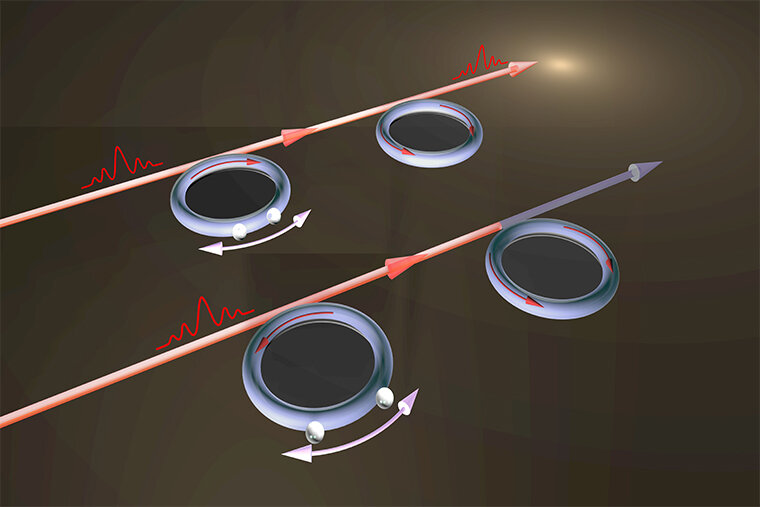
For the experiment, researchers directed light toward a pair of indirectly coupled resonators from the left (see illustration). The lightwave entered the first resonator, which was "tuned" to ensure light traveled clockwise. The light bounced around the perimeter, then exited, continuing along the fiber to the second, lower-quality resonator.
There, the light was scattered by the resonator's imperfections and some of it began traveling counter clockwise along the perimeter. The light wave then returned to the fiber, but headed back toward the first resonator.
Critically, researchers not only used the nanoparticle in the first resonator to make the lightwaves move clockwise, they also tuned it in a way that, as the light waves propagated back and forth between resonators, a special interference pattern would form. As a result of that pattern, the light in the resonators was cancelled out, so to speak, allowing the light traveling along the fiber to eek by, rendering the system transparent.
It would be as if someone shined a light on a brick wall -- no light would get through. But then another person with another flashlight shined it in the same spot and, all of a sudden, that spot in the wall became transparent.
One of the more important -- and interesting -- functions of EIT is its ability to create "slow light." The speed of light is always constant, but the actual value of that speed can change based on the properties of the medium through which it moves. In a vacuum, light always travels at 300,000,000 meters per second.
With EIT, people have slowed light down to leight meters per second, Wang said. "That can have significant influence on the storage of light information. If light is slowed down, we have enough time to use the encoded information for optical quantum computing or optical communication." If engineers can better control EIT, they can more reliably depend on slow light for these applications.
Manipulating EIT could also be used in the development of long distance communication. A tuning resonator can be indirectly coupled to another resonator kilometers away along the same fiber optic cable. "You could change the transmitted light down the line," Yang said.
This could be critical for, among other things, quantum encryption.
The research team also included collaborators at Yale University, University of Chicago and the University of Southern California.

Story Source:
Materials provided by Washington University in St. Louis. Original written by Brandie Jefferson. Note: Content may be edited for style and length.

Thursday, January 9, 2020

Researchers build a particle accelerator that fits on a chip

Subatomic particle collisions illustration (stock image).

On a hillside above Stanford University, the SLAC National Accelerator Laboratory operates a scientific instrument nearly 2 miles long. In this giant accelerator, a stream of electrons flows through a vacuum pipe, as bursts of microwave radiation nudge the particles ever-faster forward until their velocity approaches the speed of light, creating a powerful beam that scientists from around the world use to probe the atomic and molecular structures of inorganic and biological materials.
Now, for the first time, scientists at Stanford and SLAC have created a silicon chip that can accelerate electrons -- albeit at a fraction of the velocity of that massive instrument -- using an infrared laser to deliver, in less than a hair's width, the sort of energy boost that takes microwaves many feet.
Writing in the Jan. 3 issue of Science, a team led by electrical engineer Jelena Vuckovic explained how they carved a nanoscale channel out of silicon, sealed it in a vacuum and sent electrons through this cavity while pulses of infrared light -- to which silicon is as transparent as glass is to visible light -- were transmitted by the channel walls to speed the electrons along.
The accelerator-on-a-chip demonstrated in Science is just a prototype, but Vuckovic said its design and fabrication techniques can be scaled up to deliver particle beams accelerated enough to perform cutting-edge experiments in chemistry, materials science and biological discovery that don't require the power of a massive accelerator.
"The largest accelerators are like powerful telescopes. There are only a few in the world and scientists must come to places like SLAC to use them," Vuckovic said. "We want to miniaturize accelerator technology in a way that makes it a more accessible research tool."
Team members liken their approach to the way that computing evolved from the mainframe to the smaller but still useful PC. Accelerator-on-a-chip technology could also lead to new cancer radiation therapies, said physicist Robert Byer, a co-author of the Science paper. Again, it's a matter of size. Today, medical X-ray machines fill a room and deliver a beam of radiation that's tough to focus on tumors, requiring patients to wear lead shields to minimize collateral damage.
"In this paper we begin to show how it might be possible to deliver electron beam radiation directly to a tumor, leaving healthy tissue unaffected," said Byer, who leads the Accelerator on a Chip International Program, or ACHIP, a broader effort of which this current research is a part.
Inverse design
In their paper, Vuckovic and graduate student Neil Sapra, the first author, explain how the team built a chip that fires pulses of infrared light through silicon to hit electrons at just the right moment, and just the right angle, to move them forward just a bit faster than before.
To accomplish this, they turned the design process upside down. In a traditional accelerator, like the one at SLAC, engineers generally draft a basic design, then run simulations to physically arrange the microwave bursts to deliver the greatest possible acceleration. But microwaves measure 4 inches from peak to trough, while infrared light has a wavelength one-tenth the width of a human hair. That difference explains why infrared light can accelerate electrons in such short distances compared to microwaves. But this also means that the chip's physical features must be 100,000 times smaller than the copper structures in a traditional accelerator. This demands a new approach to engineering based on silicon integrated photonics and lithography.
Vuckovic's team solved the problem using inverse design algorithms that her lab has developed. These algorithms allowed the researchers to work backward, by specifying how much light energy they wanted the chip to deliver, and tasking the software with suggesting how to build the right nanoscale structures required to bring the photons into proper contact with the flow of electrons.
"Sometimes, inverse designs can produce solutions that a human engineer might not have thought of," said R. Joel England, a SLAC staff scientist and co-author on the Science paper.
The design algorithm came up with a chip layout that seems almost otherworldly. Imagine nanoscale mesas, separated by a channel, etched out of silicon. Electrons flowing through the channel run a gantlet of silicon wires, poking through the canyon wall at strategic locations. Each time the laser pulses -- which it does 100,000 times a second -- a burst of photons hits a bunch of electrons, accelerating them forward. All of this occurs in less than a hair's width, on the surface of a vacuum-sealed silicon chip, made by team members at Stanford.
The researchers want to accelerate electrons to 94 percent of the speed of light, or 1 million electron volts (1MeV), to create a particle flow powerful enough for research or medical purposes. This prototype chip provides only a single stage of acceleration, and the electron flow would have to pass through around 1,000 of these stages to achieve 1MeV. But that's not as daunting at it may seem, said Vuckovic, because this prototype accelerator-on-a-chip is a fully integrated circuit. That means all of the critical functions needed to create acceleration are built right into the chip, and increasing its capabilities should be reasonably straightforward.
The researchers plan to pack a thousand stages of acceleration into roughly an inch of chip space by the end of 2020 to reach their 1MeV target. Although that would be an important milestone, such a device would still pale in power alongside the capabilities of the SLAC research accelerator, which can generate energy levels 30,000 times greater than 1MeV. But Byer believes that, just as transistors eventually replaced vacuum tubes in electronics, light-based devices will one day challenge the capabilities of microwave-driven accelerators.
Meanwhile, in anticipation of developing a 1MeV accelerator on a chip, electrical engineer Olav Solgaard, a co-author on the paper, has already begun work on a possible cancer-fighting application. Today, highly energized electrons aren't used for radiation therapy because they would burn the skin. Solgaard is working on a way to channel high-energy electrons from a chip-sized accelerator through a catheter-like vacuum tube that could be inserted below the skin, right alongside a tumor, using the particle beam to administer radiation therapy surgically.
"We can derive medical benefits from the miniaturization of accelerator technology in addition to the research applications," Solgaard said.

Story Source:
Materials provided by Stanford University. Original written by Tom Abate. Note: Content may be edited for style and length.

Tuesday, December 24, 2019

Bioengineer Researchers Discover New Type of Cell Communication

Collective intercellular communication through ultra-fast hydrodynamic trigger waves:
Researchers studying one of the longest single cell organisms—Spirostomum ambiguum—which can grow up to lengths of 4mm (a unicellular organism observable to the naked eye) have discovered that it is also one of the fastest cells ever documented. The gargantuan protist can contract its long body by 60% within milliseconds, experiencing an acceleration force of up to 14g.

The contractile behavior protects the unicellular organism from would-be predators, as small vacuoles along the cellular membrane containing toxins are dispersed when undergoing the extreme g forces of the contraction. Remarkably, researchers have discovered that the contractions also generate long-ranged vortex flows that function as hydrodynamic signals to other Spirostomum.
This is the first time that hydrodynamic cellular signaling has been documented, and opens the door to investigating other instances where cells are communicating via long-range vorticular hydrodynamic signals.

RSF—in perspective:

RSF—in perspective: the subcellular components that allow for this blazingly fast contractile motion are microtubules. Microtubules are a central facet of current theories of quantum consciousness and cellular information processing producing memory and intelligent behavior.
As such, it is perhaps not so surprising that the intelligent behavior and communication via hydrodynamic signaling from rapid contraction by this unicellular organism involves microtubules.

Monday, December 23, 2019

Researchers discover brain circuit linked to food impulsivity

Eating popcorn at a movie (stock image).
Credit: © Zoriana / Adobe Stock

You're on a diet, but the aroma of popcorn in the movie theater lobby triggers a seemingly irresistible craving.
Within seconds, you've ordered a tub of the stuff and have eaten several handfuls.
Impulsivity, or responding without thinking about the consequences of an action, has been linked to excessive food intake, binge eating, weight gain and obesity, along with several psychiatric disorders including drug addiction and excessive gambling.
A team of researchers that includes a faculty member at the University of Georgia has now identified a specific circuit in the brain that alters food impulsivity, creating the possibility scientists can someday develop therapeutics to address overeating.
The team's findings were published recently in the journal Nature Communications.
"There's underlying physiology in your brain that is regulating your capacity to say no to (impulsive eating)," said Emily Noble, an assistant professor in the UGA College of Family and Consumer Sciences who served as lead author on the paper. "In experimental models, you can activate that circuitry and get a specific behavioral response."
Using a rat model, researchers focused on a subset of brain cells that produce a type of transmitter in the hypothalamus called melanin concentrating hormone (MCH).
While previous research has shown that elevating MCH levels in the brain can increase food intake, this study is the first to show that MCH also plays a role in impulsive behavior, Noble said.
"We found that when we activate the cells in the brain that produce MCH, animals become more impulsive in their behavior around food," Noble said.
To test impulsivity, researchers trained rats to press a lever to receive a "delicious, high-fat, high-sugar" pellet, Noble said. However, the rat had to wait 20 seconds between lever presses. If the rat pressed the lever too soon, it had to wait an additional 20 seconds.
Researchers then used advanced techniques to activate a specific MCH neural pathway from the hypothalamus to the hippocampus, a part of the brain involved with learning and memory function.
Results indicated MCH doesn't affect how much the animals liked the food or how hard they were willing to work for the food. Rather, the circuit acted on the animals' inhibitory control, or their ability to stop themselves from trying to get the food."Activating this specific pathway of MCH neurons increased impulsive behavior without affecting normal eating for caloric need or motivation to consume delicious food," Noble said. "Understanding that this circuit, which selectively affects food impulsivity, exists opens the door to the possibility that one day we might be able to develop therapeutics for overeating that help people stick to a diet without reducing normal appetite or making delicious foods less delicious."

Story Source:
Materials provided by University of Georgia. Original written by Cal Powell. Note: Content may be edited for style and length.

Saturday, December 21, 2019

Researchers determine age for last known settlement by a direct ancestor to modern humans

Homo erectus skull (stock image).
Credit: © stockdevil / Adobe Stock

Homo erectus, one of modern humans' direct ancestors, was a wandering bunch. After the species dispersed from Africa about two million years ago, it colonized the ancient world, which included Asia and possibly Europe.
But about 400,000 years ago, Homo erectus essentially vanished. The lone exception was a spot called Ngandong, on the Indonesian island of Java. But scientists were unable to agree on a precise time period for the site -- until now.
In a new study published in the journal Nature, an international team of researchers led by the University of Iowa; Macquarie University; and the Institute of Technology Bandung, Indonesia, dates the last existence of Homo erectus at Ngandong between 108,000 and 117,000 years ago.
The researchers time-stamped the site by dating animal fossils from the same bonebed where 12 Homo erectus skull caps and two tibia had been found, and then dated the surrounding land forms -- mostly terraces below and above Ngandong -- to establish an accurate record for the primeval humans' possible last stand on Earth.
"This site is the last known appearance of Homo erectus found anywhere in the world," says Russell Ciochon, professor in the Department of Anthropology at Iowa and co-corresponding author on the study. "We can't say we dated the extinction, but we dated the last occurrence of it. We have no evidence Homo erectus lived later than that anywhere else."
The research team presents 52 new age estimates for the Ngandong evidence. They include animal fossil fragments and sediment from the rediscovered fossil bed where the original Homo erectus remains were found by Dutch surveyors in the 1930s, and a sequence of dates for the river terraces below and above the fossil site.
In addition, the researchers determined when mountains south of Ngandong first rose by dating stalagmites from caves in the Southern Mountains. This allowed them to determine when the Solo River began coursing through the Ngandong site, and the river terrace sequence was created.
"You have this incredible array of dates that are all consistent," Ciochon says. "This has to be the right range. That's why it's such a nice, tight paper. The dating is very consistent."
"The issues with the dating of Ngandong could only ever be resolved by an appreciation of the wider landscape," says Kira Westaway, associate professor at Macquarie University and a joint-lead author on the paper. "Fossils are the byproducts of complex landscape processes. We were able to nail the age of the site because we constrained the fossils within the river deposit, the river terrace, the sequence of terraces, and the volcanically active landscape."
Previous research by Ciochon and others shows Homo erectus hopscotched its way across the Indonesian archipelago, and arrived on the island of Java about 1.6 million years ago. The timing was good: The area around Ngandong was mostly grassland, the same environment that cradled the species in Africa. Plants and animals were abundant. While the species continued to venture to other islands, Java, it appears, likely remained home -- or least a way station -- to some bands of the species.
However, around 130,000 years ago, the environment at Ngandong changed, and so did Homo erectus's fortunes.
"There was a change in climate," Ciochon explains. "We know the fauna changed from open country, grassland, to a tropical rainforest (extending southward from today's Malaysia). Those were not the plants and animals that Homo erectus was used to, and the species just could not adapt."
Ciochon co-led a 12-member, international team that dug at Ngandong in 2008 and in 2010, accompanied by Yan Rizal and Yahdi Zaim, the lead researchers from the Institute of Technology, Bandung, on the excavation. Using notes from the Dutch surveyors' excavation in the 1930s, the team found the original Homo erectus bone bed at Ngandong and re-exposed it, collecting and dating 867 animal fossil fragments. Meanwhile, Westaway's team had been dating the surrounding landscapes, such as the terraces, during that time.
"It was coincidental" the teams were working in the same place -- one group at the fossil bed, the other group dating the surrounding area, Ciochon says.
"With the data we had, we couldn't really date the Ngandong fossils," Ciochon continues. "We had dates on them, but they were minimum ages. So, we couldn't really say how old, although we knew we were in the ballpark. By working with Kira, who had vast amount of dating data for the terraces, mountains, and other landscape features, we were able to provide precise regional chronological and geomorphic contexts for the Ngandong site."
Researchers from multiple institutions contributed to the manuscript, including those from the Institute of Technology in Bandung, Indonesia; the University of Wollongong, Australia; the University of Texas-Austin; Griffith University in Nathan, Australia; Southern Cross University in Lismore, Australia; the University of Oxford, United Kingdom; the Geological Agency in Bandung; the University of Queensland in Brisbane Australia; the University of New England in Armidale, Australia; the University of Copenhagen in Denmark; Minnesota State University-Mankato; Bluestone Heights in Cleveland, Ohio; the University of Alberta in Edmonton, Canada; Rutgers University; Indiana University; and Illinois State University.
Rizal is a joint-lead author on the paper. E. Arthur Bettis III, emeritus professor in the Department of Earth and Environmental Sciences at Iowa, is a contributing author.
Funders include the University of Iowa, the Australian Research Council, the Wenner-Gren Foundation for Anthropological Research, the Geological Survey Institute in Bandung, and the Villum Foundation.

Story Source:
Materials provided by University of Iowa. Original written by Richard C. Lewis. Note: Content may be edited for style and length.

Tuesday, December 17, 2019

Researchers reconstruct spoken words as processed in nonhuman primate brains

Rhesus macaque

A team of Brown University researchers has used a brain-computer interface to reconstruct English words from neural signals recorded in the brains of nonhuman primates. The research, published in the journal Nature Communications Biology, could be a step toward developing brain implants that may help people with hearing loss, the researchers say.
"What we've done is to record the complex patterns of neural excitation in the secondary auditory cortex associated with primates' hearing specific words," said Arto Nurmikko, a professor in Brown's School of Engineering, a research associate in Brown's Carney Institute for Brain Science and senior author of the study. "We then use that neural data to reconstruct the sound of those words with high fidelity.
"The overarching goal is to better understand how sound is processed in the primate brain," Nurmikko added, "which could ultimately lead to new types of neural prosthetics."
The brain systems involved in the initial processing of sound are similar in humans and non-human primates. The first level of processing, which happens in what's called the primary auditory cortex, sorts sounds according to attributes like pitch or tone. The signal then moves to the secondary auditory cortex, where it's processed further. When someone is listening to spoken words, for example, this is where the sounds are classified by phonemes -- the simplest features that enable us to distinguish one word from another. After that, the information is sent to other parts of the brain for the processing that enables human comprehension of speech.
But because that early-stage processing of sound is similar in humans and non-human primates, learning how primates process the words they hear is useful, even though they likely don't understand what those words mean.
For the study, two pea-sized implants with 96-channel microelectrode arrays recorded the activity of neurons while rhesus macaques listened to recordings of individual English words and macaque calls. In this case, the macaques heard fairly simple one- or two-syllable words -- "tree," "good," "north," "cricket" and "program."
The researchers processed the neural recordings using computer algorithms specifically developed to recognize neural patterns associated with particular words. From there, the neural data could be translated back into computer-generated speech. Finally, the team used several metrics to evaluate how closely the reconstructed speech matched the original spoken word that the macaque heard. The research showed the recorded neural data produced high-fidelity reconstructions that were clear to a human listener.
The use of multielectrode arrays to record such complex auditory information was a first, the researchers say.
"Previously, work had gathered data from the secondary auditory cortex with single electrodes, but as far as we know this is the first multielectrode recording from this part of the brain," Nurmikko said. "Essentially we have nearly 200 microscopic listening posts that can give us the richness and higher resolution of data which is required."
One of the goals of the study, for which doctoral student Jihun Lee led the experiments, was to test whether any particular decoding model algorithm performed better than others. The research, in collaboration with Wilson Truccolo, a computational neuroscience expert, showed that recurrent neural networks (RNNs) -- a type of machine learning algorithm often used in computerized language translation -- produced the highest-fidelity reconstructions. The RNNs substantially outperformed more traditional algorithms that have been shown to be effective in decoding neural data from other parts of the brain.
Christopher Heelan, a research associate at Brown and co-lead author of the study, thinks the success of the RNNs comes from their flexibility, which is important in decoding complex auditory information.
"More traditional algorithms used for neural decoding make strong assumptions about how the brain encodes information, and that limits the ability of those algorithms to model the neural data," said Heelan, who developed the computational toolkit for the study. "Neural networks make weaker assumptions and have more parameters allowing them to learn complicated relationships between the neural data and the experimental task."
Ultimately, the researchers hope, this kind of research could aid in developing neural implants the may aid in restoring peoples' hearing.
"The aspirational scenario is that we develop systems that bypass much of the auditory apparatus and go directly into the brain," Nurmikko said. "The same microelectrodes we used to record neural activity in this study may one day be used to deliver small amounts of electrical current in patterns that give people the perception of having heard specific sounds."
The research was supported by the U.S. Defense Advanced Research Projects Agency (N66001-17-C-4013) and a private gift to Brown.

Story Source:
Materials provided by Brown UniversityNote: Content may be edited for style and length.

Sunday, December 15, 2019

Researchers discover brain circuit linked to food impulsivity

Eating popcorn at a movie 

You're on a diet, but the aroma of popcorn in the movie theater lobby triggers a seemingly irresistible craving.
Within seconds, you've ordered a tub of the stuff and have eaten several handfuls.
Impulsivity, or responding without thinking about the consequences of an action, has been linked to excessive food intake, binge eating, weight gain and obesity, along with several psychiatric disorders including drug addiction and excessive gambling.
A team of researchers that includes a faculty member at the University of Georgia has now identified a specific circuit in the brain that alters food impulsivity, creating the possibility scientists can someday develop therapeutics to address overeating.
The team's findings were published recently in the journal Nature Communications.
"There's underlying physiology in your brain that is regulating your capacity to say no to (impulsive eating)," said Emily Noble, an assistant professor in the UGA College of Family and Consumer Sciences who served as lead author on the paper. "In experimental models, you can activate that circuitry and get a specific behavioral response."
Using a rat model, researchers focused on a subset of brain cells that produce a type of transmitter in the hypothalamus called melanin concentrating hormone (MCH).
While previous research has shown that elevating MCH levels in the brain can increase food intake, this study is the first to show that MCH also plays a role in impulsive behavior, Noble said.
"We found that when we activate the cells in the brain that produce MCH, animals become more impulsive in their behavior around food," Noble said.
To test impulsivity, researchers trained rats to press a lever to receive a "delicious, high-fat, high-sugar" pellet, Noble said. However, the rat had to wait 20 seconds between lever presses. If the rat pressed the lever too soon, it had to wait an additional 20 seconds.
Researchers then used advanced techniques to activate a specific MCH neural pathway from the hypothalamus to the hippocampus, a part of the brain involved with learning and memory function.
Results indicated MCH doesn't affect how much the animals liked the food or how hard they were willing to work for the food. Rather, the circuit acted on the animals' inhibitory control, or their ability to stop themselves from trying to get the food."Activating this specific pathway of MCH neurons increased impulsive behavior without affecting normal eating for caloric need or motivation to consume delicious food," Noble said. "Understanding that this circuit, which selectively affects food impulsivity, exists opens the door to the possibility that one day we might be able to develop therapeutics for overeating that help people stick to a diet without reducing normal appetite or making delicious foods less delicious."

Story Source:
Materials provided by University of Georgia. Original written by Cal Powell. Note: Content may be edited for style and length.